. 2006 Jun 15;107(12):4898-906.
doi: 10.1182/blood-2005-09-3596. Epub 2006 Feb 21.

Clarifying the role of Stat5 in lymphoid development and Abelson-induced transformation

Affiliations

Clarifying the role of Stat5 in lymphoid development and Abelson-induced transformation

Andrea Hoelbl et al. Blood. .

Abstract

The Stat5 transcription factors Stat5a and Stat5b have been implicated in lymphoid development and transformation. Most studies have employed Stat5a/b-deficient mice where gene targeting disrupted the first protein-coding exon, resulting in the expression of N-terminally truncated forms of Stat5a/b (Stat5a/b(DeltaN/DeltaN) mice). We have now reanalyzed lymphoid development in Stat5a/b(null/null) mice having a complete deletion of the Stat5a/b gene locus. The few surviving Stat5a/b(null/null) mice lacked CD8(+) T lymphocytes. A massive reduction of CD8(+) T cells was also found in Stat5a/b(fl/fl) lck-cre transgenic animals. While gammadelta T-cell receptor-positive (gammadeltaTCR(+)) cells were expressed at normal levels in Stat5a/b(DeltaN/DeltaN) mice, they were completely absent in Stat5a/b(null/null) animals. Moreover, B-cell maturation was abrogated at the pre-pro-B-cell stage in Stat5a/b(null/null) mice, whereas Stat5a/b(DeltaN/DeltaN) B-lymphoid cells developed to the early pro-B-cell stage. In vitro assays using fetal liver-cell cultures confirmed this observation. Most strikingly, Stat5a/b(null/null) cells were resistant to transformation and leukemia development induced by Abelson oncogenes, whereas Stat5a/b(DeltaN/DeltaN)-derived cells readily transformed. These findings show distinct lymphoid defects for Stat5a/b(DeltaN/DeltaN) and Stat5a/b(null/null) mice and define a novel functional role for the N-termini of Stat5a/b in B-lymphoid transformation.


Figures
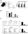
Figure 1
Figure 1. Impaired CD8+ and γδ T-cell development in Stat5a/bnull/null survivor mice
(A) Picture of a Stat5a/bnull/null survivor mouse (left) compared with a Stat5a/b+/+ littermate at the age of 4 weeks. Representative body weight values and numbers of Stat5a/bnull/null mice and littermates surviving up to day 8, weaning (day 21), or day 42 are depicted. (B) Numbers of CD4/CD8/γδTCR+ T lymphocytes in thymi of Stat5a/bnull/null, Stat5a/bΔN/ΔN, and respective littermate controls. (C) Flow cytometric analysis of CD4+, CD8+, and CD4+/CD8+ cells in thymi and lymph nodes of 5 Stat5a/bnull/null survivors and 2 individual Stat5a/bnull/+ and 2 Stat5a/b+/+ littermate controls. Data are summarized in bar graphs. Due to the small size of thymi and lymph nodes, cells were pooled and did therefore not allow generation of error bars (B-C).
Figure 2
Figure 2. Impaired CD8+ T-cell development in Stat5a/bf/f lck-cre mice
(A) Splenic cells of 3 individual Stat5a/bf/f lck-cre (nos. 1-3) and 2 Stat5a/bf/f (nos. 4-5) mice were magnetically sorted for Thy1.2+ cells, and Stat5a/b expression was assessed by Western blot analysis. (B) Representative flow cytometric profile of CD4+, CD8+, and CD4+/CD8+ cells in thymus, blood, spleen, and lymph nodes of Stat5a/bf/f lck-cre mice and Stat5a/bf/f controls. Asterisks denote significant difference as determined by a paired t test. (C) Analysis of transcriptional expression of pim-1, CIS, and cyclin D2 genes by semiquantitative RT-PCR. Splenic T cells were stimulated for 4 hours with α-CD3 (1 μg/mL) and human IL-2 (hIL-2; 1000 U/mL) to induce Stat5a/b target gene transcription. (D) Schematic model for the role of Stat5a/b in T-cell developmental choices. Stages affected in Stat5a/bnull/null survivor mice and/or Stat5a/bf/f lck-cre mice are indicated. The time point of Cre-recombinase activation under the control of the distal lck promoter is also depicted.
Figure 3
Figure 3. B-cell development is arrested at the pre–pro-B-cell stage in Stat5a/bnull/null survivor mice
(A) Percentages of pre–pro-B, early pro-B, and late pro-B cells in bone marrow and (B) mature B cells in spleen and lymph node of 5 Stat5a/bnull/null survivors compared with 2 Stat5a/bnull/+ and 2 Stat5a/b+/+ controls. Due to the small body size, bone marrows were pooled and therefore did not allow generation of error bars. (C) Schematic model for maturation of B-cell developmental fractions A-F. As indicated, individual maturational stages were distinguished by differential surface expression of B220, CD43, CD19, BP-1, IgM, and IgD. The different blocks in Stat5a/bΔN/ΔN and Stat5a/bnull/null mice are indicated by vertical lines. CLP indicates common lymphoid progenitor.
Figure 4
Figure 4. B-cell development is arrested at the pre–pro-B-cell stage in Stat5a/bnull/null fetal liver–derived cultures
Fetal livers of 2 embryos of each genotype were pooled (ED 14) and cocultivated on an OP-9 fibroblast feeder layer in the presence of IL-7, Flt-3L, and SCF (10 ng/mL each). Outgrowth of pro-B-cell stages (fractions A-C), immature (fraction E), and mature (fraction F) B cells over a 12-day period is depicted.
Figure 5
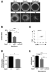
Figure 5. Abelson-induced transformation is dependent on Stat5a/b in vitro
(A) Ab-MuLV–induced colony formation of Stat5a/b+/+, Stat5a/bnull/+, and Stat5a/bnull/null fetal liver cells in methylcellulose. Single-colony pictures of each phenotype are depicted in the bottom panels. Stat5a/bnull/null cells showed no ability to form growth factor–independent colonies. (B) Summary of data obtained from Ab-MuLV–induced colony formation assays represent means ± SEM of 4 embryos per genotype (each performed in triplicates). (C) Surface expression of B-lineage markers was verified by flow cytometric analysis (right; data of one representative CD19+ CD43+ colony is shown). (D) [3H]thymidine incorporation of fetal liver–derived Stat5a/b+/+ and Stat5a/bnull/+ Ab-MuLV–transformed cell lines. Stat5a/b-deficient fetal livers did not give rise to stable transformed cell lines. Data represent means ± SEM of 2 cell lines per genotype. cpm indicates counts per minute. (E) Ab-MuLV–induced colony formation of Stat5a/b+/+ (n = 2), Stat5a/bnull/+ (n = 2), and Stat5a/bnull/null (n = 5; pooled) survivor bone marrow cells in methylcellulose. Stat5a/bnull/null survivor cells showed no ability to form growth factor–free colonies. Experiment was performed in triplicates. Asterisks denote significant differences as determined by a one-way ANOVA followed by a Tukey test (A,C) or a paired t test (B).
Figure 6
Figure 6. Abelson-induced transformation is dependent on Stat5 in vivo
(A) Kaplan-Meier plot of Rag2−/− mice that received a transplant of either Stat5a/b+/+, Stat5a/bnull/+, or a 4:1 mixture of Stat5a/bnull/null and Stat5a/b+/+ freshly Ab-MuLV–transformed bone marrow cells (5 mice/group; 1 × 10⁶ cells each mouse). Genotyping PCR analysis of mice used for 4:1 mixture is depicted. wt indicates wild type. (B) Immunoblotting for Stat5a/b of leukemic cells derived from bone marrow of Rag2−/− mice that received a transplant of either Stat5a/b+/+ or a 4:1 mixture of Stat5a/bnull/null/Stat5a/b+/+ bone marrow. (C) PCR analysis of ex vivo–derived cell lines. Representative data of bone marrow (BM)–, peripheral blood (PB)–, spleen-, and lymph node (LN)–derived leukemic cell lines of one Rag2−/− mouse that received a transplant of a 4:1 mixture of Stat5a/bnull/null/Stat5a/b+/+ bone marrow cells, which was later killed. All cultures derived from Rag2−/− mice that received transplants of Stat5a/bnull/null/Stat5a/b+/+ cells were Stat5a/b+/+ as determined by PCR analysis.

Similar articles

Cited by

References

    1. Calo V, Migliavacca M, Bazan V, et al. STAT proteins: from normal control of cellular events to tumorigenesis. J Cell Physiol. 2003;197:157–168. - PubMed
    1. Levy DE, Darnell JE., Jr. Stats: transcriptional control and biological impact. Nat Rev Mol Cell Biol. 2002;3:651–662. - PubMed
    1. Murray R. Physiologic roles of interleukin-2, interleukin-4, and interleukin-7. Curr Opin Hematol. 1996;3:230–234. - PubMed
    1. Moriggl R, Topham DJ, Teglund S, et al. Stat5 is required for IL-2-induced cell cycle progression of peripheral T cells. Immunity. 1999;10:249–259. - PubMed
    1. Gouilleux-Gruart V, Debierre-Grockiego F, Gouilleux F, et al. Activated Stat related transcription factors in acute leukemia. Leuk Lymphoma. 1997;28:83–88. - PubMed

Publication types

MeSH terms

Substances