

Pulsed high-intensity focused ultrasound enhances thrombolysis in an in vitro model
- PMID: 16493016
- PMCID: PMC2386885
- DOI: 10.1148/radiol.2391042181
Pulsed high-intensity focused ultrasound enhances thrombolysis in an in vitro model
Abstract
Purpose: To evaluate the use of pulsed high-intensity focused ultrasound exposures to improve tissue plasminogen activator (tPA)-mediated thrombolysis in an in vitro model.
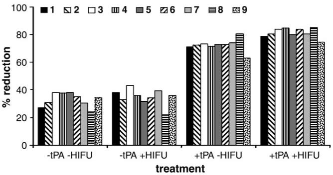
Materials and methods: All experimental work was compliant with institutional guidelines and HIPAA. Clots were formed by placing 1 mL of human blood in closed-off sections of pediatric Penrose tubes. Four experimental groups were evaluated: control (nontreated) clots, clots treated with pulsed high-intensity focused ultrasound only, clots treated with tPA only, and clots treated with pulsed high-intensity focused ultrasound plus tPA. The focused ultrasound exposures (real or sham) were followed by incubations of the clots in tPA with saline or in saline only. Thrombolysis was measured as the relative reduction in the mass of the clot. D-Dimer assays also were performed. Two additional experiments were performed and yielded dose-response curves for two exposure parameters: number of pulses per raster point and total acoustic power. Radiation force-induced displacements caused by focused ultrasound exposures were simulated in the clots. A Tukey-Kramer honestly significant difference test was performed for comparisons between all pairs of experimental groups.
Results: The clots treated with focused ultrasound alone did not show significant increases in thrombolysis compared with the control clots. The clots treated with focused ultrasound plus tPA showed a 50% ([30.2/20.1]/20.1) increase in the degree of thrombolysis compared with the clots treated with tPA only (P < .001), further corroborating the d-dimer assay results (P < .001). Additional experiments revealed how increasing both the number of pulses per raster point and the total acoustic power yielded corresponding increases in the thrombolysis rate. In the latter experiment, simulations performed at a range of power settings revealed a direct correlation between increased displacement and observed thrombolysis rate.
Conclusion: The rate of tPA-mediated thrombolysis can be enhanced by using pulsed high-intensity focused ultrasound exposure in vitro.
(c) RSNA, 2006.
Figures


References
-
- De Wet CJ, Pear RG. Postoperative thrombotic complications: venous thromboembolism—deep-vein thrombosis and pulmonary embolism. Anesthesiol Clin North Am. 1999;17:895–922.
-
- Goldhaber S. Epidemiology of pulmonary embolism and deep vein thrombosis. In: Bloom AL, Forbes CD, Thombas DP, Tuddenham EGD, editors. Haemostasis and thrombosis. 3rd ed. Churchill Livingstone; New York, NY: 1994. pp. 1327–1333.
-
- a National Institutes of Health Consensus Development Conference Thombolytic therapy in thrombosis. Ann Intern Med. 1980;93:141–144. - PubMed
-
- Tachibana K. Enhancement of fibrinolysis with ultrasound energy. J Vasc Interv Radiol. 1992;3:299–303. - PubMed
-
- Luo H, Steffen W, Cercek B, Arunasalam S, Maurer G, Siegel RJ. Enhancement of thrombolysis by external ultrasound. Am Heart J. 1993;125:1564–1569. - PubMed
Publication types
MeSH terms
Grants and funding
LinkOut - more resources
Full Text Sources
Medical
