

Muscle-associated triglyceride measured by computed tomography and magnetic resonance spectroscopy
- PMID: 16493125
- PMCID: PMC2677802
- DOI: 10.1038/oby.2006.10
Muscle-associated triglyceride measured by computed tomography and magnetic resonance spectroscopy
Abstract
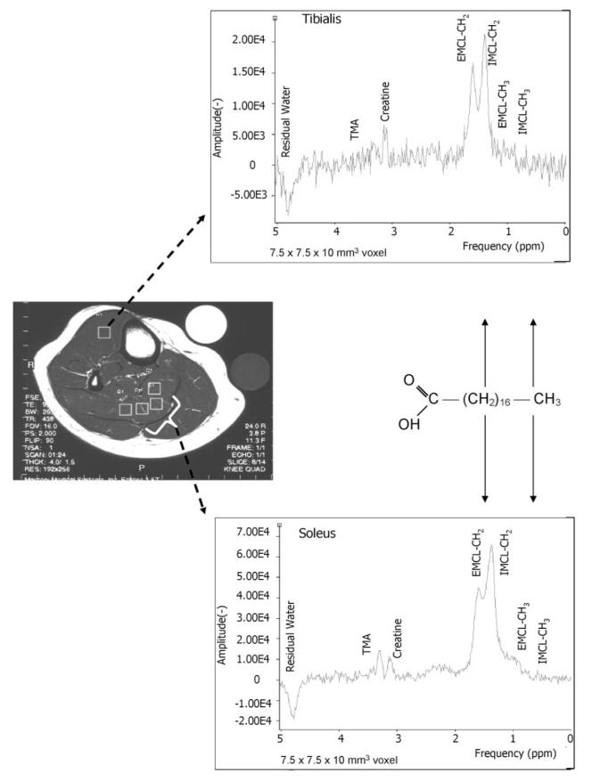
Objective: Muscle triglyceride can be assessed in vivo using computed tomography (CT) and 1H magnetic resonance spectroscopy (MRS), two techniques that are based on entirely different biophysical principles. Little is known, however, about the cross-correlation between these techniques and their test-retest reliability.
Research methods and procedures: We compared mean muscle attenuation (MA) in soleus and tibialis anterior (TA) muscles measured by CT with intra- and extramyocellular lipids (IMCL and EMCL, respectively) measured by MRS in 51 volunteers (26 to 72 years of age, BMI = 25.5 to 39.3 kg/m2). MA of midthighs was also measured in a subset (n = 19). Test-retest measurements were performed by CT (n = 6) and MRS (n = 10) in separate sets of volunteers.
Results: MA of soleus was significantly associated with IMCL (r = -0.64) and EMCL, which by multiple regression analysis was explained mostly by IMCL (p < 0.001) rather than EMCL (beta = -0.010, p = 0.94). Muscle triglyceride was lower in TA than in soleus, and MA of TA was significantly correlated with EMCL (r = -0.40) but not IMCL (r = -0.16). By CT, MA of midthighs was correlated with MA in soleus (r = 0.40, p = 0.07) and whole calf (r = 0.62, p < 0.05). Finally, both MA and IMCL were highly reliable in soleus (coefficient of variation = < 2% and 6.7%, respectively) and less reliable in TA (4% and 10%, respectively).
Discussion: These results support the use of both CT and MRS as reliable methods for assessing skeletal muscle lipid.
Figures


References
-
- Goodpaster BH, Kelley DE. Role of muscle in triglyceride metabolism. Curr Opin Lipidol. 1998;9:231–6. - PubMed
-
- Kelley DE, Goodpaster BH. Skeletal muscle triglyceride. An aspect of regional adiposity and insulin resistance. Diabetes Care. 2001;24:933–41. - PubMed
-
- Pan D, Lillioja S, Kriketos A, et al. Skeletal muscle triglyceride levels are inversely related to insulin action. Diabetes. 1997;46:983–8. - PubMed
-
- Goodpaster BH, He J, Watkins S, Kelley DE. Skeletal muscle lipid content and insulin resistance: evidence for a paradox in endurance-trained athletes. J Clin Endocrinol Metab. 2001;86:5755–61. - PubMed
-
- Goodpaster BH, Katsiaras A, Kelley DE. Enhanced fat oxidation through physical activity is associated with improvements in insulin sensitivity in obesity. Diabetes. 2003;52:2191–7. - PubMed
