. 2006 Jun 1;396(2):381-9.
doi: 10.1042/BJ20051878.

Engineered covalent leucotoxin heterodimers form functional pores: insights into S-F interactions

Affiliations

Engineered covalent leucotoxin heterodimers form functional pores: insights into S-F interactions

Olivier Joubert et al. Biochem J. .

Abstract

The staphylococcal alpha-toxin and bipartite leucotoxins belong to a single family of pore-forming toxins that are rich in beta-strands, although the stoichiometry and electrophysiological characteristics of their pores are different. The different known structures show a common beta-sandwich domain that plays a key role in subunit-subunit interactions, which could be targeted to inhibit oligomerization of these toxins. We used several cysteine mutants of both HlgA (gamma-haemolysin A) and HlgB (gamma-haemolysin B) to challenge 20 heterodimers linked by disulphide bridges. A new strategy was developed in order to obtain a good yield for S-S bond formation and dimer stabilization. Functions of the pores formed by 14 purified dimers were investigated on model membranes, i.e. planar lipid bilayers and large unilamellar vesicles, and on target cells, i.e. rabbit and human red blood cells and polymorphonuclear neutrophils. We observed that dimers HlgA T28C-HlgB N156C and HlgA T21C-HlgB T157C form pores with similar characteristics as the wild-type toxin, thus suggesting that the mutated residues are facing one another, allowing pore formation. Our results also confirm the octameric stoichiometry of the leucotoxin pores, as well as the parity of the two monomers in the pore. Correctly assembled heterodimers thus constitute the minimal functional unit of leucotoxins. We propose amino acids involved in interactions at one of the two interfaces for an assembled leucotoxin.


Figures
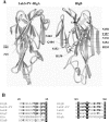
Figure 1
Figure 1. Basic three-dimensional structures and sequence alignment of leucotoxins
(A) Three-dimensional structures of LukS-PV (PDB code 1T5R; right-rear view) that might be comparable with that of HlgA and HlgB (PDB code 1LKF; left-rear view) and the location of the cysteine-substituted amino acids. Amino acids thought to interact each other are underlined and in italics respectively. (B) Sequence alignment of the S. aureus Hlg (GenBank® accession number X81586), PVL (GenBank® accession number X72700), LukE and LukD (GenBank® accession number Y13225) and α-toxin (Hla, GenBank® accession number M90536). Conserved residues are shown in white on a black background, while common residues to bipartite leucotoxins are shown in bold; numbering of the amino acids is given on the basis of HlgA and HlgB respectively.
Figure 2
Figure 2. Engineering and purification of the Hlg covalent heterodimer HlgA T28C–HlgB N156C
SDS/10–15% PAGE and silver staining. Lane 1, molecular ladder; lanes 2 and 6, HlgA T28C (0.3 μg) and HlgB N156C (0.5 μg) respectively, under reducing conditions; lanes 3 and 5, HlgA T28C (1 μg) and HlgB N156C (0.8 μg) respectively under oxidizing conditions; lane 4, HlgA T28C–HlgB N156C (1 μg of the coupling reaction); lane 7, mixture of recombinant WT HlgA (0.2 μg) and WT HlgB (0.15 μg); lane 8, FPLC-purified HlgA T28C–HlgB N156C. Molecular-mass sizes are given in kDa.
Figure 3
Figure 3. HlgA–HlgB heterodimers induce variable haemolytic activities in HRBCs compared with the WT toxin
HRBCs were incubated in the presence of 20 nM heterodimers obtained by combining HlgA T21C (A) or HlgA T28C (B). The haemolytic activity was monitored by following the decrease in absorbance at 650 nm. Kinetics were then normalized as a function of the 100% of haemolysis obtained for the control, as described in the Materials and methods section. Dimers are abbreviated to A (HlgA) or B (HlgB) and the positions of residues that were mutated to cysteine.
Figure 4
Figure 4. Pore-forming activity and ethidium bromide entry into human PMNs are induced by the HlgA–HlgB heterodimers
Human PMNs were incubated in the presence of 20 nM heterodimers obtained by combining HlgA T21C (A) or HlgA T28C (B), and formation of pores was followed by the entry of ethidium bromide and its combination with nucleic acids, as described in the Materials and methods section. The WT HlgA–HlgB leucotoxin, applied at 20 nM, was used as a control. Dimers are abbreviated to A (HlgA) or B (HlgB) and the positions of residues that were mutated to cysteine.
Figure 5
Figure 5. HlgA–HlgB heterodimers are responsible for different Ca2+ influxes into human PMNs as evaluated by flow cytometry
(A) Each heterodimer and the control (HlgA plus HlgB) were used at 20 nM. Dimers HlgA T21C–HlgB N158C and HlgA T28C–HlgB R155C activities were similar to those of HlgA T21C–HlgB T157C and HlgA T28C–HlgB N156C. Dimers HlgA T21C–HlgB Q104C, HlgA T21C–HlgB R155C and HlgA T21C–HlgB N156C activities were intermediate, similar to those of HlgA T21C–HlgB S154C and HlgA T28C–HlgB S154C. Dimer HlgA T28C–HlgB Q104C activity was null like that of HlgA T21C–HlgB N103C. Dimers are abbreviated to A (HlgA) or B (HlgB) and the positions of residues that were mutated to cysteine. (B) Compared calcium entries induced by all purified heterodimers, from serial data as in (A). +++, similarly as active as the control (fluorescence greater than 70% of the control at 5 min incubation); ++, intermediate activity (fluorescence between 20 and 70% of the control at 5 min incubation); +, weak activity (fluorescence less than 20% of the control at 5 min incubation and greater than 10% at 15 min of incubation); −, no calcium entry.
Figure 6
Figure 6. Formation of ion channels in planar lipid bilayers by Hlg and dimers
Representative stepwise current increases corresponding to the opening of single ion channels of Hlg WT and all of the active heterodimers as indicated on the left of each trace. Each protein was added to the cis side at the concentrations reported in brackets of Table 2. The applied voltage was +40 mV in all cases. The height of each step was used to calculate the conductance of that pore as reported in Table 2.
Figure 7
Figure 7. HlgA and HlgB and heterodimer oligomers formed in solution or after their insertion into human PMN membranes
Oligomers were analysed by SDS/3–8% (w/v) PAGE and were revealed by immunoblotting with anti-LukS-PV and anti-LukF-PV affinity-purified rabbit antibodies with WT. Lane 1, PMNs only; lane 2, HlgA and HlgB (0.2 ng each) were mixed for 1 h at room temperature and analysed; lane 3, 4 ng of each HlgA and HlgB was concatemerized in solution with 0.3 mM glutaraldehyde; lane 4, same experiment as in lane 3, but with 3 mM glutaraldehyde; lane 5, HlgA and HlgB were applied to human PMNs and retrieved after saponin treatment, but without glutaraldehyde; lane 6, HlgA (15 ng) and HlgB (15 ng) oligomers were applied to human PMNs and retrieved after saponin and glutaraldehyde treatments, before being electrophoresed without boiling; lanes 7 and 8, temperature, 0 °C or 23 °C respectively, of the saponin treatment did not affect the retrieving of oligomers that were boiled after the glutaraldehyde treatment; lanes 9–11, 30 ng of heterodimers HlgA T28C–HlgB N156C, HlgA T28C–HlgB T157C and HlgA T21C–HlgB R155C respectively were applied to human PMNs, and oligomers were prepared as in lane 8 and show octamers, hexamers, tetramers and dimers as well as monomers in minor quantities. Any heterodimers with full or medium activities produced similar oligomers (not shown); lane 12, HlgA T28C–N103C (or HlgA T28C–Q104C, not shown) failed to promote oligomers under the same conditions as in lanes 7 to 11; lanes 13, 14 and 15, oligomers obtained from other active heterodimers: HlgA T21C–HlgB N156C, HlgA T21C–HlgB T157C and HlgA T21C–HlgB N158C. Molecular-mass sizes are given in kDa.

Similar articles

Cited by

References

    1. Prévost G., Mourey L., Colin D. A., Monteil H., Dalla Serra M., Menestrina G. α-Toxin and β-barrel pore-forming toxins (leucocidins, α-, γ-, and δ-cytolysins) of Staphylococcus aureus. In: Alouf J. E., Popoff M. R., editors. The Comprehensive Sourcebook of Bacterial Protein Toxins. London: Academic Press; 2005. pp. 588–605.
    1. Rossjohn J., Feil S. C., McKinstry W. J., Tweten R. K., Parker M. W. Structure of a cholesterol-binding, thiol-activated cytolysin and a model of its membrane form. Cell. 1997;89:685–692. - PubMed
    1. Moayeri M., Leppla S. H. The roles of anthrax toxin in pathogenesis. Curr. Opin. Microbiol. 2004;7:19–24. - PubMed
    1. Aktories K. Clostridial ADP-ribosylating toxins: effects on ATP and GTP-binding proteins. Mol. Cell. Biochem. 1994;138:167–176. - PubMed
    1. Thelestam M., Olofsson A., Blomqvist L., Hebert H. Oligomerization of cell-bound staphylococcal α-toxin in relation to membrane permeabilisation. Biochim. Biophys. Acta. 1991;1062:245–254. - PubMed

Publication types

MeSH terms