Comparative Study
. 2006 Mar;50(3):899-909.
doi: 10.1128/AAC.50.3.899-909.2006.

Preclinical profile of VX-950, a potent, selective, and orally bioavailable inhibitor of hepatitis C virus NS3-4A serine protease

Affiliations
Comparative Study

Preclinical profile of VX-950, a potent, selective, and orally bioavailable inhibitor of hepatitis C virus NS3-4A serine protease

Robert B Perni et al. Antimicrob Agents Chemother. 2006 Mar.

Abstract

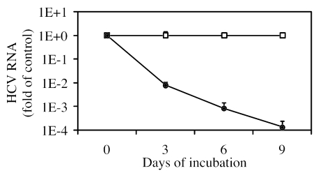
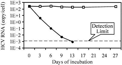
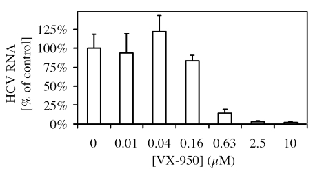
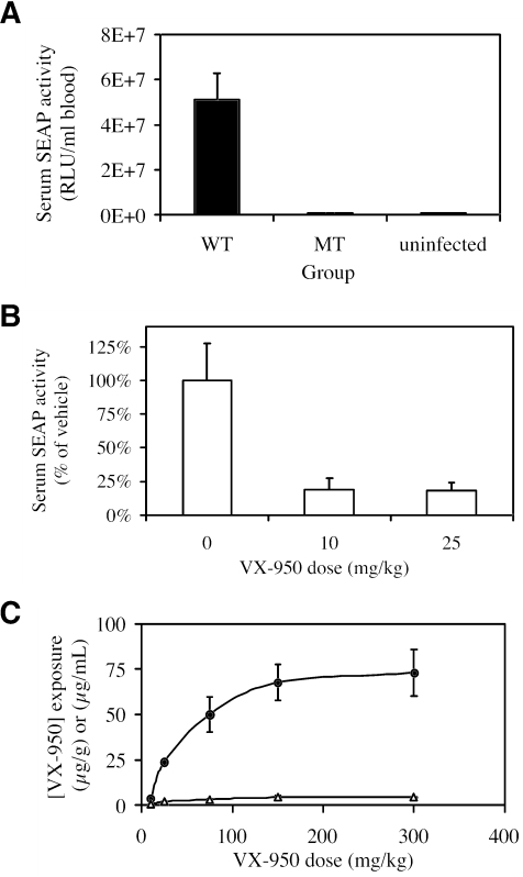
VX-950 is a potent, selective, peptidomimetic inhibitor of the hepatitis C virus (HCV) NS3-4A serine protease, and it demonstrated excellent antiviral activity both in genotype 1b HCV replicon cells (50% inhibitory concentration [IC50] = 354 nM) and in human fetal hepatocytes infected with genotype 1a HCV-positive patient sera (IC50 = 280 nM). VX-950 forms a covalent but reversible complex with the genotype 1a HCV NS3-4A protease in a slow-on, slow-off process with a steady-state inhibition constant (K(i)*) of 7 nM. Dissociation of the covalent enzyme-inhibitor complex of VX-950 and genotype 1a HCV protease has a half-life of almost an hour. A >4-log10 reduction in the HCV RNA levels was observed after a 2-week incubation of replicon cells with VX-950, with no rebound of viral RNA observed after withdrawal of the inhibitor. In several animal species, VX-950 exhibits a favorable pharmacokinetic profile with high exposure in the liver. In a recently developed HCV protease mouse model, VX-950 showed excellent inhibition of HCV NS3-4A protease activity in the liver. Therefore, the overall preclinical profile of VX-950 supports its candidacy as a novel oral therapy against hepatitis C.


Figures

FIG. 1.
FIG. 1.
(A) Chemical diagrams of the natural NS5A/5B substrate (compound 1), with its P6 to P4′ residues and the cleavage bond (indicated by solid arrow), and the HCV NS3-4A protease inhibitor VX-950 (compound 2), with the bond attacked by Ser139 of HCV protease (indicated by a solid arrow) and two carbonyl groups (indicated by an open arrow). (B) Schematic representation of the covalent EI* complex structure based on an X-ray co-crystal structure of the HCV NS3-4A protease and VX-950. The inhibitor is shown at the left and the protease at the right. A covalent but reversible bond is formed between the hydroxyl group of catalytic Ser139 of the HCV protease and the keto-carbonyl group of VX-950. In addition, two hydrogen bonds (indicated by dashed lines) are formed between two main chain amide NH groups of the protease (Gly137 and Ser139) and the amide carbonyl group of VX-950.
FIG. 2.
FIG. 2.
Concentration-dependent reduction of HCV replicon RNA levels by VX-950. HCV replicon cells were incubated with various concentrations of VX-950 for 48 h. At the end of the 48-h incubation, total cellular RNA was extracted by RNeasy-96. The level of HCV replicon RNA remaining was then determined by QRT-PCR, as described in Materials and Methods, and was shown as a percentage of replicon RNA levels in control cells incubated with 0.5% DMSO. Each bar represents the average of results from 5 cell culture replicates with the SD. Data for one representative IC50 determination is shown.
FIG. 3.
FIG. 3.
Nine-day HCV replicon clearance assay with VX-950. The cells were incubated with 0.2% DMSO (open square) or 7 μM VX-950 (filled circle) for 3, 6, or 9 days. Fresh 0.2% DMSO or 7 μM VX-950 in medium was added to the cells every 3 days. At the end of each incubation period, cell numbers were determined by the tetrazolium-based cell viability assay based on an established standard curve, and the level of HCV RNA in the cells was determined by the QRT-PCR assay. The copy number of HCV RNA per cell in each sample is plotted as relative (on a log scale) to that in replicon cells incubated with 0.2% DMSO (control) for the same period of time. Each datum point represents the average of the results from 5 cell culture replicates with the SD.
FIG. 4.
FIG. 4.
Lack of rebound of HCV replicon RNA after a 13-day incubation with VX-950. HCV replicon cells were incubated with 0.2% DMSO (open square) or 17.5 μM VX-950 (filled circle) in the absence of G418. Cells were split every 3 to 4 days, and fresh 0.2% DMSO or 17.5 μM VX-950 in medium was added to the cells. After 13 days of incubation, VX-950 was withdrawn and 0.25 mg/ml G418 was added to enrich the remaining HCV replicon-positive cells that are capable of growing in the presence of G418 (rebound). The cultures were monitored for another 14 days in the presence of G418. The cell number was determined by the Guava ViaCount assay, as described in Materials and Methods, and the level of HCV RNA in the cells was determined by the QRT-PCR assay. The absolute numbers of HCV replicon RNA copies per viable cell are shown. The detection limit of the QRT-PCR assay is indicated by a dashed line.
FIG. 5.
FIG. 5.
Concentration-dependent reduction of HCV RNA in infected primary fetal hepatocytes by VX-950. The primary human fetal hepatocytes were isolated from a fetal liver and plated as described in Materials and Methods. Serum from an HCV-infected patient was used to infect the primary fetal hepatocytes 5 days after the plating. The inoculum was removed 24 h later, and the cells were incubated with VX-950 for 5 days. At the end of the 5-day incubation, the cells were washed and total cellular RNA was extracted by RNeasy-96. The level of HCV replicon RNA remaining was then determined by QRT-PCR, as described in Materials and Methods, and was shown as a percentage of the level of replicon RNA in cells incubated with 0.5% DMSO (control). Each bar represents the average of the results from 4 to 6 cell culture replicates with the SD.
FIG. 6.
FIG. 6.
VX-950 inhibits HCV NS3-4A protease and reduces SEAP levels in the NS3-4A protease mouse model. (A) Three groups of SCID mice (n = 6 per group) were given an injection of recombinant adenovirus Ad-WT-HCVpro-SEAP (WT) or Ad-MT-HCVpro-SEAP (MT) or not given an adenovirus injection (uninfected) through the tail vein. Serum collected at 24 h after injection was diluted fivefold with water, and SEAP activity was determined using the Phospha-Light detection system. Each datum point represents the average (±SEM) of SEAP activity per ml of undiluted serum in each dosing group of 6 mice. (B) Three groups of SCID mice were injected with recombinant adenovirus Ad-WT-HCVpro-SEAP through the tail vein. Two of these groups (n = 6 per group) were given 2 oral doses of VX-950 (10 or 25 mg/kg per administration): the first was given 2 h before the virus injection and the second was given 10 h after injection. The third group (n = 10) was dosed with vehicle only. Serum collected at 24 h postinjection was diluted, and the SEAP activity was determined as described above. The average (±SEM) SEAP activity per ml of undiluted serum in each dose group is plotted as the percentage of that of the vehicle group. (C) Six groups of SCID mice (n = 6 per group) were given a single oral administration of vehicle (0) or VX-950 (10, 25, 75, 150, or 300 mg/kg). The serum and liver samples were collected 1 h postdosing (shown here) from half of each dose group (n = 3) or 12 h postdosing (not shown) from the other half of each dose group (n = 3). The average VX-950 concentration at 1 h postinjection, determined by the chiral LC/MS/MS separation method, is plotted for the serum (open triangle, in μg/ml) or the liver (filled circle, in μg/g) along with SD.

References

    1. Castet, V., C. Fournier, A. Soulier, R. Brillet, J. Coste, D. Larrey, D. Dhumeaux, P. Maurel, and J. M. Pawlotsky. 2002. Alpha interferon inhibits hepatitis C virus replication in primary human hepatocytes infected in vitro. J. Virol. 76:8189-8199. - PMC - PubMed
    1. De Francesco, R., L. Tomei, S. Altamura, V. Summa, and G. Migliaccio. 2003. Approaching a new era for hepatitis C virus therapy: inhibitors of the NS3-4A serine protease and the NS5B RNA-dependent RNA polymerase. Antivir. Res. 58:1-16. - PubMed
    1. Di Marco, S., M. Rizzi, C. Volpari, M. A. Walsh, F. Narjes, S. Colarusso, R. De Francesco, V. G. Matassa, and M. Sollazzo. 2000. Inhibition of the hepatitis C virus NS3/4A protease. The crystal structures of two protease-inhibitor complexes. J. Biol. Chem. 275:7152-7157. - PubMed
    1. Fried, M. W., M. L. Shiffman, K. R. Reddy, C. Smith, G. Marinos, F. L. Goncales, Jr., D. Haussinger, M. Diago, G. Carosi, D. Dhumeaux, A. Craxi, A. Lin, J. Hoffman, and J. Yu. 2002. Peginterferon alfa-2a plus ribavirin for chronic hepatitis C virus infection. N. Engl. J. Med. 347:975-982. - PubMed
    1. Grakoui, A., D. W. McCourt, C. Wychowski, S. M. Feinstone, and C. M. Rice. 1993. Characterization of the hepatitis C virus-encoded serine proteinase: determination of proteinase-dependent polyprotein cleavage sites. J. Virol. 67:2832-2843. - PMC - PubMed

Publication types

MeSH terms

LinkOut - more resources