. 2006 Mar;50(3):1013-20.
doi: 10.1128/AAC.50.3.1013-1020.2006.

SCH 503034, a mechanism-based inhibitor of hepatitis C virus NS3 protease, suppresses polyprotein maturation and enhances the antiviral activity of alpha interferon in replicon cells

Affiliations

SCH 503034, a mechanism-based inhibitor of hepatitis C virus NS3 protease, suppresses polyprotein maturation and enhances the antiviral activity of alpha interferon in replicon cells

B A Malcolm et al. Antimicrob Agents Chemother. 2006 Mar.

Abstract

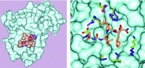
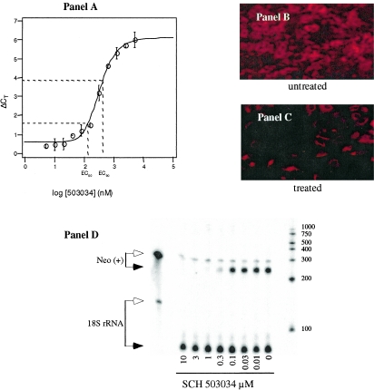
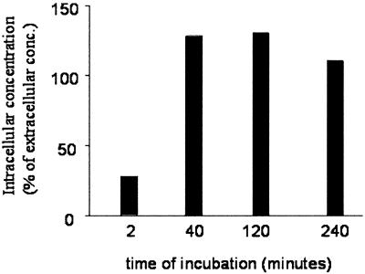
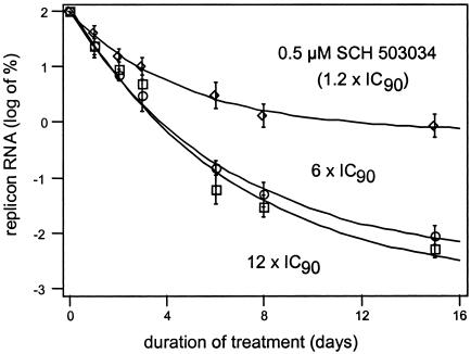
Cleavage of the hepatitis C virus (HCV) polyprotein by the viral NS3 protease releases functional viral proteins essential for viral replication. Recent studies by Foy and coworkers strongly suggest that NS3-mediated cleavage of host factors may abrogate cellular response to alpha interferon (IFN-alpha) (E. Foy, K. Li, R. Sumpter, Jr., Y.-M. Loo, C. L. Johnson, C. Wang, P. M. Fish, M. Yoneyama, T. Fujita, S. M. Lemon, and M. Gale, Jr., Proc. Natl. Acad. Sci. USA 102:2986-2991, 2005, and E. Foy, K. Li, C. Wang, R. Sumpter, Jr., M. Ikeda, S. M. Lemon, and M. Gale, Jr., Science 300:1145-1148, 2003). Blockage of NS3 protease activity therefore is expected to inhibit HCV replication by both direct suppression of viral protein production as well as by restoring host responsiveness to IFN. Using structure-assisted design, a ketoamide inhibitor, SCH 503034, was generated which demonstrated potent (overall inhibition constant, 14 nM) time-dependent inhibition of the NS3 protease in cell-free enzyme assays as well as robust in vitro activity in the HCV replicon system, as monitored by immunofluorescence and real-time PCR analysis. Continuous exposure of replicon-bearing cell lines to six times the 90% effective concentration of SCH 503034 for 15 days resulted in a greater than 4-log reduction in replicon RNA. The combination of SCH 503034 with IFN was more effective in suppressing replicon synthesis than either compound alone, supporting the suggestion of Foy and coworkers that combinations of IFN with protease inhibitors would lead to enhanced therapeutic efficacy.


Figures

FIG. 1.
FIG. 1.
SCH 503034 structure and chemical name.
FIG. 2.
FIG. 2.
Progress curve of peptide hydrolysis by the single-chain HCV protease domain (genotype 1b) showing time-dependent inhibition by SCH 503034. SCH 503034 was incubated with the single-chain genotype 1b NS3 protease (see Materials and Methods). Reactions were initiated by addition of enzyme and absorbance (abs) was monitored for 120 min. Data were fitted to the two-step “slow-binding” inhibition model to obtain kinetic parameters.
FIG. 3.
FIG. 3.
SCH 503034 complexed with the HCV NS3 protease. A. Connolly surface for the NS3 protease. SCH 503034 is rendered in CPK format (Corey-Pauling-Koltun space-filling model): gold, carbon; red, oxygen; and blue, nitrogen. B. A close up of the NS3:SCH 503034 complex showing side chains that are perturbed upon binding of SCH 503034. Color code for SCH 503034 is the same as that for panel A. Side chains of the complex are colored as follows: carbon, green; nitrogen, blue; and oxygen, red. Side chains of the apoenzyme structure are shown in purple.
FIG. 4.
FIG. 4.
Effect of SCH 503034 on HCV replicon RNA synthesis in Huh-7 cells. A. Real-time PCR (Taqman) analysis of the HCV replicon RNA level relative to the GAPDH internal control (ΔCT). Huh-7 cells were treated with increasing concentrations of SCH 503034 for 72 h in the presence of G418. Increases in ΔCT (y axis) indicate decreasing replicon RNA levels; each ΔCT reflects a twofold change in RNA level from baseline. A representative set of data is shown. Immunofluorescent detection of NS3 protein expression following exposure to SCH 503034. HCV replicon-bearing Huh-7 cells were treated with vehicle alone (0.5% DMSO) (B) or with SCH 503034 (1× EC90) (C) for 72 h and then probed with anti-NS3 polyclonal antibodies and visualized by staining with an immunofluorescently labeled secondary antibody. A comparable number of cells are present in each field. D. Detection of replicon RNA from drug-treated cells by RNase protection assay. Clone 16 cell cultures were treated with SCH 503034 for 3 days as described in Materials and Methods. Cell monolayers were recovered in cell lysis buffer and processed for RPAs detecting positive-strand replicon genomic RNA (via the Neo gene) and 18S rRNA (internal control). RPA products were separated on a TBE-5% polyacrylamide gel electrophoresis gel containing urea. Arrows show the positions of digested (filled) and undigested (open) riboprobe. Similar results were obtained in several experiments using other replicon cell lines (data not shown).
FIG. 5.
FIG. 5.
Inhibition of polyprotein processing in HCV replicon cells. Replicon cells were treated with SCH 503034 (0 to 100 μM, 1:3 serial dilution). Cells were harvested at various time points as indicated, and cell lysates were analyzed by Western blot for NS5A as described in Materials and Methods. The position of the high-molecular-weight processing intermediate (possibly NS3-NS5B) is indicated by an arrow. Additional processing intermediates, detected at 6 h posttreatment, are indicated by a bracket.
FIG. 6.
FIG. 6.
Uptake of [14C]SCH 503034 by replicon-bearing cells. Primary human hepatocytes were incubated in the presence of 10 μM radiolabeled SCH 503034 for various time intervals as indicated. Cells were harvested, washed, and counted. The intracellular concentration of SCH 503034 was estimated and compared with the initial concentration in the media. Results are expressed as percentages of medium concentration (conc.).
FIG. 7.
FIG. 7.
Effect of treatment duration and inhibitor concentration on replicon RNA level. Replicon-bearing Huh-7 cells (clone 16) were incubated for 14 days in media (without G418 selection) containing 0.5 μM (1.2 × 90% inhibitory concentration), 2.5 μM (6 × EC90) SCH 503034, or 5 μM (12 × EC90) SCH 503034. Compound and medium were refreshed daily. HCV replicon RNA level was determined by TaqMan as described in Materials and Methods and normalized to time zero. Experiments were performed in triplicate, and error bars represent the standard deviations.
FIG. 8.
FIG. 8.
Effect of SCH 503034 on alpha interferon (IFN) potency in vitro. Replicon cells were treated simultaneously with SCH 503034 and IFN as described in Materials and Methods. A. Nonparametric (spline-fit) analysis of efficacy as measured by the reduction in HCV replicon RNA. Axes are normalized to maximum dose. B. The isobole at the 90% suppression level. The line of additivity is shown. Although the isobole trends below the line of additivity, the deflection is within the error of the assay and therefore indicates simple additivity of alpha interferon and SCH 503034. IC90, 90% inhibitory concentration.

References

    1. Alter, H. J., and L. B. Seeff. 2000. Recovery, persistence, and sequelae in hepatitis C virus infection: a perspective on long-term outcome. Semin. Liver Dis. 20:17-35. - PubMed
    1. Bain, V. G. 2001. Effect of HCV viral dynamics on treatment design: lessons learned from HIV. Am. J. Gastroenterol. 96:2818-2828. - PubMed
    1. Bartenschlager, R., L. Ahlborn-Laake, J. Mous, and H. Jacobsen. 1993. Nonstructural protein 3 of the hepatitis C virus encodes a serine-type proteinase required for cleavage at the NS3/4 and NS4/5 junctions. J. Virol. 67:3835-3844. - PMC - PubMed
    1. Bazan, J. F., and R. J. Fletterick. 1988. Viral cysteine proteases are homologous to the trypsin-like family of serine proteases: structural and functional implications. Proc. Natl. Acad. Sci. USA 85:7872-7876. - PMC - PubMed
    1. Berger, A., and I. Schechter. 1970. Mapping the active site of papain with the aid of peptide substrates and inhibitors. Philos. Trans. R. Soc. Lond. B. Biol. Sci. 257:249-264. - PubMed

MeSH terms

LinkOut - more resources