. 2006 Mar 14;103(11):4034-9.
doi: 10.1073/pnas.0510928103. Epub 2006 Feb 22.

MicroRNAs direct rapid deadenylation of mRNA

Affiliations

MicroRNAs direct rapid deadenylation of mRNA

Ligang Wu et al. Proc Natl Acad Sci U S A. .

Abstract

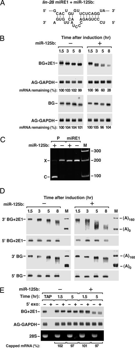
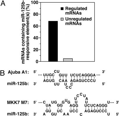
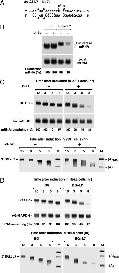
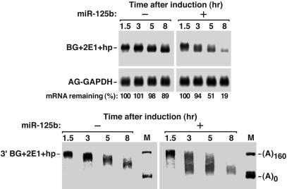
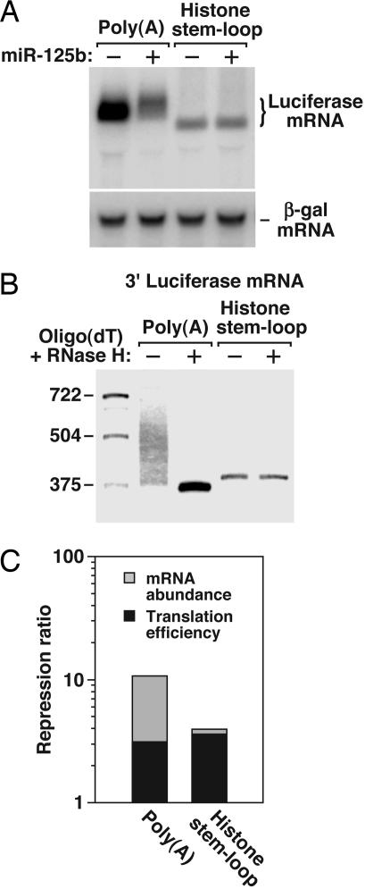
MicroRNAs (miRNAs) are ubiquitous regulators of eukaryotic gene expression. In addition to repressing translation, miRNAs can down-regulate the concentration of mRNAs that contain elements to which they are imperfectly complementary. Using miR-125b and let-7 as representative miRNAs, we show that in mammalian cells this reduction in message abundance is a consequence of accelerated deadenylation, which leads to rapid mRNA decay. The ability of miRNAs to expedite poly(A) removal does not result from decreased translation; nor does translational repression by miRNAs require a poly(A) tail, a 3' histone stem-loop being an effective substitute. These findings suggest that miRNAs use two distinct posttranscriptional mechanisms to down-regulate gene expression.


Conflict of interest statement

Conflict of interest statement: No conflicts declared.

Figures

Fig. 1.
Fig. 1.
Effect of miR-125b on the degradation of mRNA bearing lin-28 miRE1. (A) Duplex expected for lin-28 miRE1 (top) base-paired with miR-125b (bottom). (B) Decay of BG+2E1 and BG mRNA in the presence or absence of miR-125b. Total cytoplasmic RNA was extracted from transfected 293T cells at time intervals after serum stimulation to induce transient transcription of each reporter gene from its c-fos promoter. Equal amounts of each RNA sample were analyzed by electrophoresis and blotting. The relative quantity of reporter mRNA remaining at each time was calculated after normalization to AG-GAPDH mRNA (a cotransfected internal standard). (C) Inability of miR-125b to direct endonucleolytic cleavage within miRE1. Luciferase reporter mRNAs bearing miRE1 or a synthetic element (element P) perfectly complementary to miR-125b were extracted from 293T cells that did or did not produce miR-125b, ligated to an RNA oligonucleotide, and amplified by RT-PCR, using primers corresponding to sites within the ligated oligonucleotide or 0.08 kb downstream of the inserted element. The RT-PCR products were analyzed by gel electrophoresis beside DNA size markers (M). DNA sequencing confirmed that band C represented a degradation intermediate resulting from miR-125b-directed mRNA cleavage in the middle of the perfectly complementary element P. Band X, which resulted from miR-125b- independent mRNA cleavage upstream of the insertion site of miRE1 or element P, served as an internal standard; its low intensity in the leftmost lane is due to the diminished concentration of this reporter mRNA in the presence of miR-125b and competition with band C for PCR amplification. Calibration is in base pairs. (D) Deadenylation mediated by miR-125b. Equal amounts of the total cytoplasmic RNA samples examined in B were subjected to site-specific cleavage by RNase H in the presence of an oligodeoxynucleotide complementary to codons 74–81 within the coding region of BG+2E1 and BG mRNA. The 5′ and 3′ RNA fragments thereby produced were analyzed by electrophoresis and blotting, using markers (M) that corresponded in size to reporter mRNA 3′ fragments bearing no poly(A) or a 160-nt poly(A) tail. (E) Retention of the 5′ cap on mRNA undergoing deadenylation. The 1.5 and 5 h BG+2E1 mRNA samples examined in B were treated or not treated with a 5′-phosphate-dependent exonuclease and analyzed by electrophoresis and blotting. Prior treatment of the 1.5 h sample with tobacco acid pyrophosphatase (TAP) released the 5′ cap and rendered the mRNA susceptible to 5′ exonuclease digestion. 28S rRNA, which lacks a 5′ cap, served as an additional positive control for exonuclease activity. AG-GAPDH mRNA was used as a normalization standard. The percentage of BG+2E1 mRNA that was capped was calculated from the ratio of band intensities in the presence versus the absence of exonuclease treatment.
Fig. 2.
Fig. 2.
miR-125b-responsive elements in down-regulated mRNAs of P19 cells. (A) Correlation between reduced mRNA concentration and the presence of probable miR-125b-responsive elements. Mouse Genome 430A 2.0 arrays (Affymetrix) were used to compare the relative concentrations of mRNAs in undifferentiated P19 cells 24 h after mock transfection or transfection with a chemically synthesized miR-125b duplex (see Fig. 6). Two groups of mRNAs were identified whose concentration could be said with a high degree of certainty to have decreased significantly (by at least a factor of 1.40, with ≥95% confidence that the change was by at least a factor of 1.30) (see Table 1) or to have remained unchanged (≥95% confidence of a concentration change no greater than ±5%) in the presence of miR-125b. These mRNAs (22 in the first group, and 669 in the second) were then examined for 3′ UTR elements with the potential to interact productively with miR-125b [complementarity to nucleotides 2–8 or to nucleotides 1 and 3–9 of miR-125b (15, 31)], and the percentage of mRNAs with one or more such elements was calculated for each group. (B) Duplexes expected to result from base pairing of the Ajuba A1 element or the MKK7 M7 element with miR-125b.
Fig. 3.
Fig. 3.
Effect of let-7 on the deadenylation and decay of mRNA bearing the lin-28 L7 element. (A) Duplex expected for the lin-28 L7 element (top) base-paired with let-7a (bottom). (B) Influence of let-7a on the concentration of Luc+6L7 mRNA. Luciferase mRNA levels were analyzed by electrophoresis and blotting of total cytoplasmic RNA from 293T cells that had been transiently cotransfected with a luciferase reporter gene containing 0 or 6 copies of the lin-28 L7 element in its 3′ UTR, a gene encoding (+) or not encoding (−) human let-7a, and a β-galactosidase gene (internal standard). (C) Influence of let-7a on the degradation of BG+L7 mRNA in 293T cells. Analyses of mRNA deadenylation and decay similar to those in Fig. 1 were performed with RNA samples from 293T cells that had been transiently cotransfected with a BG+L7 reporter gene, a gene encoding (+) or not encoding (−) human let-7a, and a gene encoding AG-GAPDH mRNA (internal standard). (D) Deadenylation and decay of BG+L7 and BG mRNA in HeLa cells. Analyses of mRNA degradation similar to those in Fig. 1 were performed by using RNA samples from HeLa cells that had been transiently cotransfected with a reporter gene containing (BG+L7) or lacking (BG) one copy of L7 and a gene encoding AG-GAPDH mRNA (internal standard).
Fig. 4.
Fig. 4.
Accelerated deadenylation and decay in the absence of translation. Analyses of mRNA degradation similar to those in Fig. 1 were performed with RNA samples from 293T cells that had been transiently cotransfected with a modified BG+2E1 gene bearing a 40-bp inverted repeat in the 5′ UTR (BG+2E1+hp), a gene encoding (+) or not encoding (−) miR-125b, and a gene encoding AG-GAPDH mRNA (internal standard).
Fig. 5.
Fig. 5.
Translational repression in the absence of a poly(A) tail. (A) Differential effect of a 3′ histone stem-loop or a poly(A) tail on the reduction in mRNA abundance caused by miR-125b. Luciferase mRNA levels were analyzed by electrophoresis and blotting of total cytoplasmic RNA from 293T cells that had been transiently cotransfected with a gene that encoded a luciferase reporter mRNA bearing six copies of miRE1 in the 3′ UTR and ending with either a 3′ poly(A) tail (Luc+6E1) or a 3′-terminal histone H1.3 stem-loop (Luc+6E1.HSL), a gene that encoded (+) or did not encode (−) miR-125b, and a β-galactosidase gene (internal standard). (B) Confirmation of the dissimilar nature of the 3′ ends. The Luc+6E1 and Luc+6E1.HSL RNA samples from cells lacking miR-125b were also analyzed by electrophoresis and blotting after treatment, in the presence or absence of oligo(dT), with RNase H and an oligodeoxynucleotide complementary to a segment 361–386 nt upstream of the poly(A) addition site of Luc+6E1 or 376–395 nt upstream of the 3′ end of Luc+6E1.HSL (right). Calibration is in nucleotides. (C) Contributions of translation inhibition and diminished mRNA abundance to repression by miR-125b. After normalizing the concentrations of Luc+6E1 and Luc+6E1.HSL mRNA to β-galactosidase mRNA, the ratio of each in the absence or presence of miR-125b was calculated and superposed on a bar graph showing the overall degree of repression of the same reporters, as judged from relative luciferase protein levels (normalized to β-galactosidase). The effect of miR-125b on translation efficiency (black bars) corresponds to the ratio of its overall effect on protein synthesis versus its effect on mRNA concentration (gray bars). In the case of the polyadenylated reporter (Luc+6E1), which was repressed 10.7-fold, expression of miR-125b reduced translation efficiency by a factor of 3.1 ± 0.3 and mRNA abundance by a factor of 3.5 ± 0.2. In the case of the reporter ending in a histone stem-loop (Luc+6E1.HSL), which was repressed 3.9-fold, miR-125b reduced translation efficiency by a factor of 3.6 ± 0.3 and mRNA abundance by a factor of 1.1 ± 0.1.

Comment in

  • MicroRNAs: new players in an old game.
    Gupta M, Brewer G. Gupta M, et al. Proc Natl Acad Sci U S A. 2006 Mar 14;103(11):3951-2. doi: 10.1073/pnas.0601268103. Epub 2006 Mar 7. Proc Natl Acad Sci U S A. 2006. PMID: 16537465 Free PMC article. No abstract available.

Similar articles

Cited by

References

    1. Zamore P. D., Tuschl T., Sharp P. A., Bartel D. P. Cell. 2000;101:25–33. - PubMed
    1. Lee R. C., Feinbaum R. L., Ambros V. Cell. 1993;75:843–854. - PubMed
    1. Wightman B., Ha I., Ruvkun G. Cell. 1993;75:855–862. - PubMed
    1. Moss E. G., Lee R. C., Ambros V. Cell. 1997;88:637–646. - PubMed
    1. Pillai R. S., Bhattacharyya S. N., Artus C. G., Zoller T., Cougot N., Basyuk E., Bertrand E., Filipowicz W. Science. 2005;309:1573–1576. - PubMed

Publication types