Randomized Controlled Trial
. 2006 Feb 22;26(8):2260-8.
doi: 10.1523/JNEUROSCI.3386-05.2006.

Dissociating the role of ventral and dorsal premotor cortex in precision grasping

Affiliations
Randomized Controlled Trial

Dissociating the role of ventral and dorsal premotor cortex in precision grasping

Marco Davare et al. J Neurosci. .

Abstract

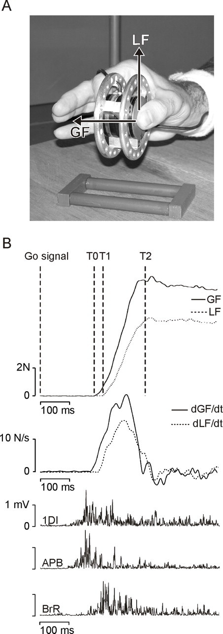
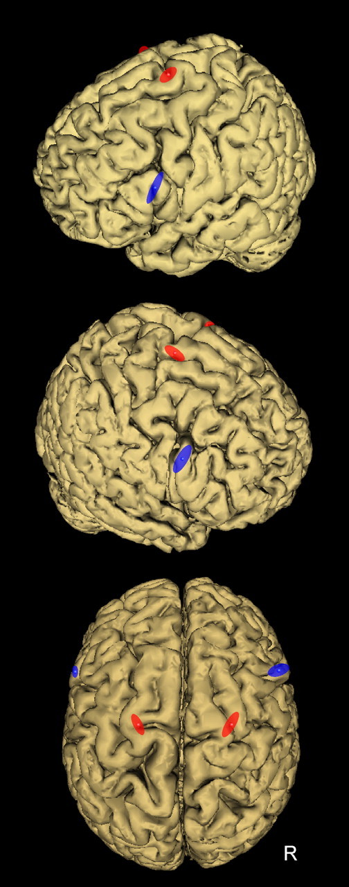
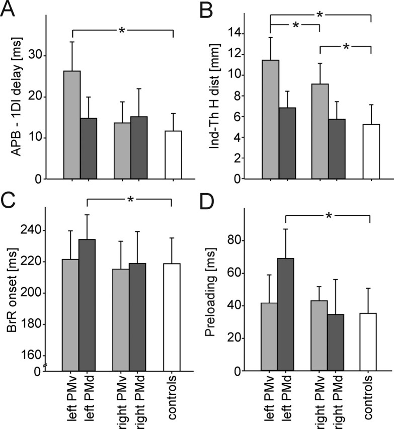
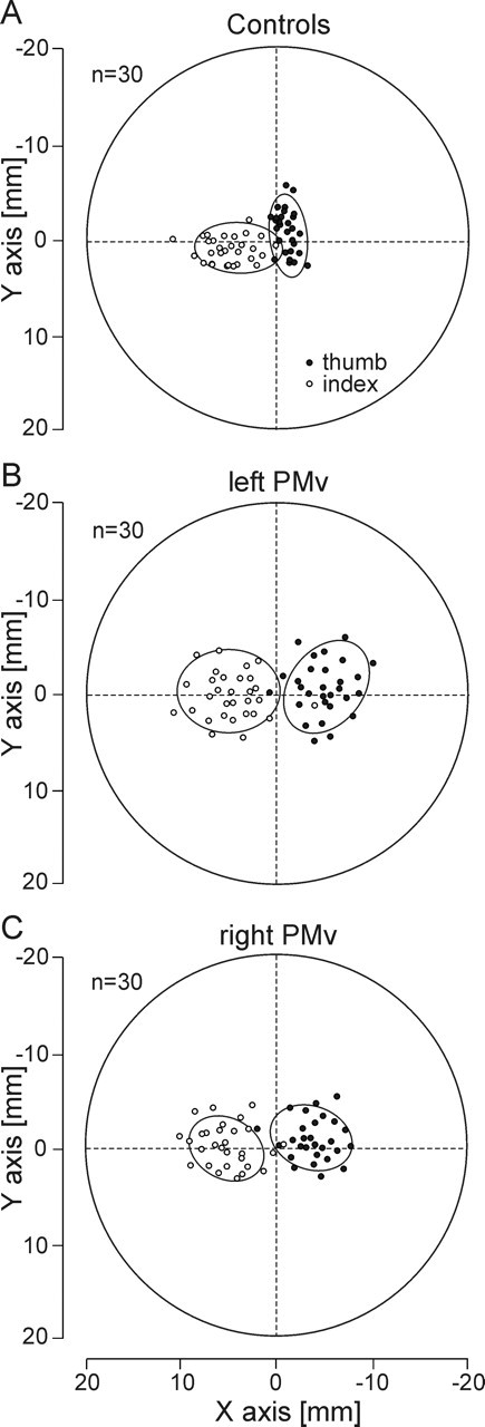
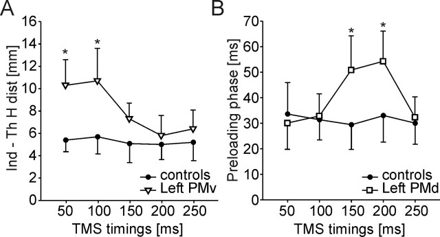
Small-object manipulation is essential in numerous human activities, although its neural bases are still essentially unknown. Recent functional imaging studies have shown that precision grasping activates a large bilateral frontoparietal network, including ventral (PMv) and dorsal (PMd) premotor areas. To dissociate the role of PMv and PMd in the control of hand and finger movements, we produced, by means of transcranial magnetic stimulation (TMS), transient virtual lesions of these two areas in both hemispheres, in healthy subjects performing a grip-lift task with their right, dominant hand. We found that a virtual lesion of PMv specifically impaired the grasping component of these movements: a lesion of either the left or right PMv altered the correct positioning of fingers on the object, a prerequisite for an efficient grasping, whereas lesioning the left, contralateral PMv disturbed the sequential recruitment of intrinsic hand muscles, all other movement parameters being unaffected by PMv lesions. Conversely, we found that a virtual lesion of the left PMd impaired the proper coupling between the grasping and lifting phases, as evidenced by the TMS-induced delay in the recruitment of proximal muscles responsible for the lifting phase; lesioning the right PMd failed to affect dominant hand movements. Finally, an analysis of the time course of these effects allowed us to demonstrate the sequential involvement of PMv and PMd in movement preparation. These results provide the first compelling evidence for a neuronal dissociation between the different phases of precision grasping in human premotor cortex.


Figures

Figure 1.
Figure 1.
The grip–lift task. A, Picture of the manipulandum used to investigate grip–lift movements. The task was always performed with the right, dominant hand, and only the thumb and index fingertips were in contact with the manipulandum. GF and LF vectors are shown for the thumb. B, Typical control grip–lift movement gathered during a sham trial. From top to bottom, GF and LF, their first derivatives (dGF/dt and dLF/dt), and the EMG activity of the 1DI, APB, and BrR. T0–T1 and T1–T2 cursors on the GF and LF traces delimit the preloading and loading phases, respectively.
Figure 2.
Figure 2.
Location of the TMS coil positions to induce virtual lesion of PMv (blue) and PMd (red). To stimulate PMv, the coil was positioned over the caudal portion of the pars opercularis of the inferior frontal gyrus (BA44), corresponding to the following normalized MNI coordinates: −60 ± 2, 16 ± 3, 23 ± 9 mm (x, y, z, mean ± SD; n = 10) and 56 ± 6, 16 ± 4, 26 ± 9 mm (n = 6) for the left and right PMv, respectively. To target PMd, the coil was positioned over the superior portion of the precentral gyrus, as delimited by the superior frontal sulcus. The mean MNI coordinates of stimulation sites for the left and right PMd were, respectively, −22 ± 3, −4 ± 4, 71 ± 4 mm (x, y, z, mean ± SD; n = 10) and 24 ± 4, −5 ± 6, 72 ± 3 mm (n = 6). Each ellipse was centered on the mean MNI coordinates of PMv and PMd stimulation points, and their surface shows the 95% confidence interval of the normalized coordinates calculated for each subject.
Figure 3.
Figure 3.
Distinctive effects of the left or right PMv or PMd inactivation on the grip–lift movement. Virtual lesions of PMv specifically altered the grasping phase of grip–lift movement, whereas lesions of the left PMd only affected the lifting phase. Histograms showing the dissociation between the effects of virtual lesions of the left and right PMv (light gray) and PMd (dark gray) when compared with the control condition (white) for four movement parameters: A, the delay between the intrinsic hand muscle recruitment, i.e., the APB and 1DI; B, the index–thumb horizontal distance (Ind-Th H dist); C, the BrR muscle onset; and D, the preloading phase duration. *p < 0.05
Figure 4.
Figure 4.
Effects of the virtual lesions of the left or right PMv on the fingertip positioning. Side view of the manipulandum showing the distribution of the fingertip positions for the thumb (black circles) and the index finger (white circles). The two graspable surfaces of the manipulandum were superimposed to represent the thumb and index fingertip positions on the same graph. The subject’s hand came from the right side of the figure. The ellipses represent the area in which 95% of the fingertip positions were found. A, Distribution of the thumb and index fingertip positions for 30 control trials in one subject. Note that the index finger contact points were always slightly farther than those of the thumb. B, A virtual lesion of the left PMv increased significantly both the horizontal distance between the thumb and index finger contact points and their variability. C, Similar effects, although less pronounced, of a virtual lesion of the right PMv on fingertip positions and distributions.
Figure 5.
Figure 5.
Distinct time course of the effects of PMv or PMd virtual lesions. A, Time course of the effect of a left PMv virtual lesion on the horizontal index–thumb distance. This parameter was found increased after left PMv stimulation but only when paired-pulse TMS was delivered 50 or 100 ms after the Go signal (white triangles) when compared with control (black circles). B, Time course of the consequence of a left PMd virtual lesion on the preloading phase duration. A virtual lesion of the left PMd led to a longer preloading phase (white squares) but only when paired-pulse TMS was delivered 150 or 200 ms when compared with controls (black circles). *p < 0.05

Comment in

References

    1. Andres M, Davare M, Pesenti M, Olivier E, Seron X (2004). Number magnitude and grip aperture interaction. NeuroReport 15:2773–2777. - PubMed
    1. Binkofski F, Buccino G (2004). Motor functions of the Broca’s region. Brain Lang 89:362–369. - PubMed
    1. Binkofski F, Buccino G, Posse S, Seitz RJ, Rizzolatti G, Freund H (1999). A fronto-parietal circuit for object manipulation in man: evidence from an fMRI-study. Eur J Neurosci 11:3276–3286. - PubMed
    1. Brochier T, Boudreau MJ, Pare M, Smith AM (1999). The effects of muscimol inactivation of small regions of motor and somatosensory cortex on independent finger movements and force control in the precision grip. Exp Brain Res 128:31–40. - PubMed
    1. Chen R, Gerloff C, Hallett M, Cohen LG (1997). Involvement of the ipsilateral motor cortex in finger movements of different complexities. Ann Neurol 41:247–254. - PubMed

Publication types