. 2006 Mar;74(3):1597-611.
doi: 10.1128/IAI.74.3.1597-1611.2006.

Binding of vitronectin by the Moraxella catarrhalis UspA2 protein interferes with late stages of the complement cascade

Affiliations

Binding of vitronectin by the Moraxella catarrhalis UspA2 protein interferes with late stages of the complement cascade

Ahmed S Attia et al. Infect Immun. 2006 Mar.

Abstract

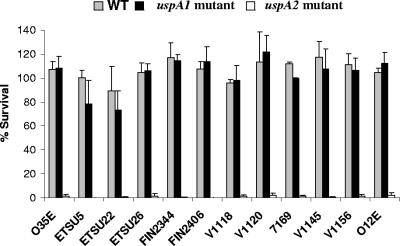
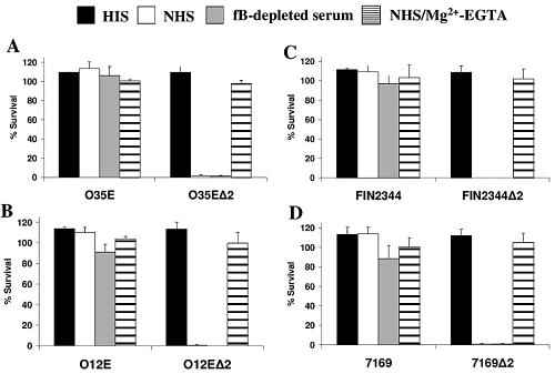
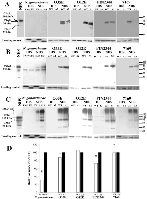
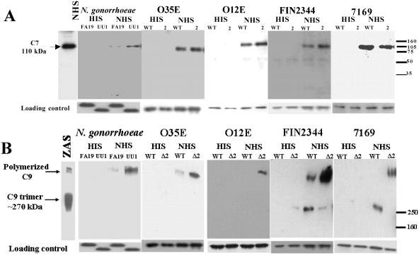
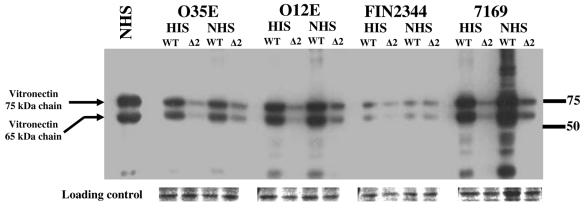
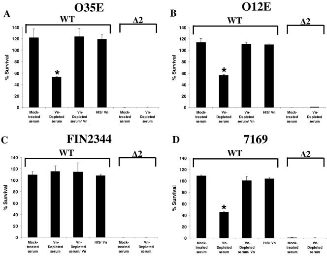
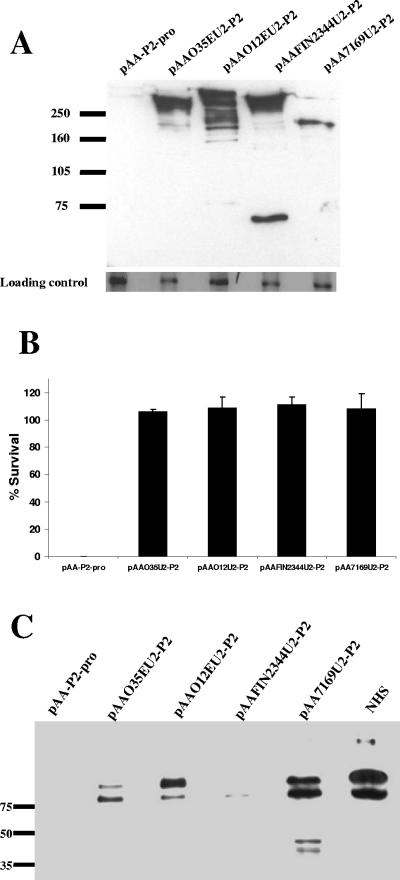
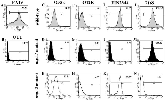
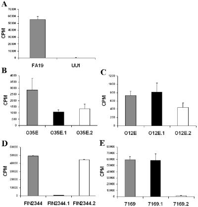
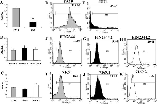
Many Moraxella catarrhalis strains are resistant to the bactericidal activity of normal human serum (NHS). The UspA2 protein of the serum-resistant strain O35E has previously been shown to be directly involved in conferring serum resistance on this strain. Testing of 11 additional serum-resistant M. catarrhalis wild-type isolates and their uspA1 and uspA2 mutants showed that the uspA1 mutants of all 11 strains were consistently serum resistant and that the uspA2 mutants of these same 11 strains were always serum sensitive. Analysis of complement deposition on four different serum-resistant M. catarrhalis strains and their serum-sensitive uspA2 mutants showed that, for three of these four strain sets, the wild-type and mutant strains bound similar amounts of early complement components. In contrast, there was a significant reduction in the amount of the polymerized C9 on the wild-type strains relative to that on the uspA2 mutants. These same three wild-type strains bound more vitronectin than did their uspA2 mutants. UspA2 proteins from these three strains, when expressed in Haemophilus influenzae, bound vitronectin and conferred serum resistance on this organism. Furthermore, vitronectin-depleted NHS exhibited bactericidal activity against these same three serum-resistant wild-type strains; addition of purified vitronectin to this serum restored serum resistance. In contrast, binding of the complement regulator C4b-binding protein by the M. catarrhalis strains used in this study was found to be highly variable and did not appear to correlate with the serum-resistant phenotype. These results indicate that binding of vitronectin by UspA2 is involved in the serum resistance of M. catarrhalis; this represents the first example of vitronectin-mediated serum resistance on a microbe.


Figures

FIG. 1.
FIG. 1.
Serum resistance of wild-type and mutant strains of M. catarrhalis. Wild-type M. catarrhalis strains and their respective uspA1 and uspA2 mutants were incubated in 10% NHS at 37°C for 30 min. Bacterial aliquots were plated at t = 0 and t = 30 min. The percentage of survival was calculated with respect to the original inoculum. These results represent the means of three independent experiments, and the error bars represent the standard deviations.
FIG. 2.
FIG. 2.
M. catarrhalis uspA2 mutants are killed via the classical pathway. Serum bactericidal assays were performed as described in Materials and Methods with four M. catarrhalis wild-type strains and their uspA2 deletion mutants (Δ2): O35E (A), O12E (B), FIN2344 (C), and 7169 (D). The four different types of sera were heat-inactivated NHS (HIS) (black bars), NHS (white bars), factor B-depleted human serum (fB) (gray bars), and NHS containing 10 mM MgCl2 and 10 mM EGTA (striped bars). These results represent the means of three independent experiments and the error bars represent the standard deviations.
FIG. 3.
FIG. 3.
Deposition of early components of the complement cascade on M. catarrhalis strains. N. gonorrhoeae strains FA19 and UU1 and M. catarrhalis wild-type (WT) strains and uspA2 mutants (Δ2) were incubated with either 10% HIS or 10% NHS for 20 min, washed three times with ice-cold GVBS, suspended in 100 μl of PBS, and then boiled with 50 μl of 3× digestion buffer. Proteins present in these samples were resolved by SDS-PAGE under reducing conditions and transferred to nitrocellulose membranes. The membranes were probed with polyclonal antibodies against C1q (the antibody recognizes mainly C1qA [29 kDa]), C1qB [26 kDa], and, to a lesser extent, C1qC [22 kDa] (A), C4 (the antibody mainly recognizes the C4β chain [75 kDa] (B), and C3 (the antibody recognizes C3α [117 kDa], C3β [75 kDa], and C3bα′ covalently bound to bacteria [C3bα′ +R] (C). As a loading control, membranes were probed with either MAb 10F3 (1) or MAb 5D2 (44); both of these MAbs recognize M. catarrhalis outer membrane antigens. For the N. gonorrhoeae samples, membranes were probed with MAb 2C3 (5). The first lanes in panels A to C contain a sample of NHS diluted 1:200 that was probed with the respective antibodies. Protein molecular mass markers (in kilodaltons) are presented on the right side of each panel. (D) Relative amounts of C3 bound to these strains as measured by flow cytometry. These experiments involved incubation of bacteria in 10% NHS for 20 min at 37°C. The cells were then placed on ice, washed three times, and subjected to flow cytometry using the same primary antibody as used in Western blot analysis. A value of 100 on the y axis represents the relative amount of C3 on the serum-sensitive strain in each pair. The data are the means from three independent experiments. The P value was 0.003 for the N. gonorrhoeae pair and 0.04 for the FIN2344 pair. For the other M. catarrhalis pairs, the differences were not significant.
FIG. 4.
FIG. 4.
Deposition of late components of the complement cascade on M. catarrhalis strains. The same whole-cell lysates described in the legend to Fig. 3 were used in this experiment. Proteins present in these samples were resolved by SDS-PAGE under nonreducing conditions. (A) Separated proteins were transferred to nitrocellulose membranes and probed with polyclonal antiserum against C7 (110 kDa); (B) separated proteins were transferred to polyvinylidene difluoride membranes and probed with a MAb against SC5b-9 which recognizes a neoepitope in polymerized C9 in the MAC. As a loading control, membranes were probed with the same antibodies as described in the legend to Fig. 3. The first lane in panel A contains a sample of NHS diluted 1:200 that was probed with the C7 antiserum. The first lane of panel B contains a control sample of ZAS that was probed with the MAb against SC5b-9 to detect polymerized C9. Protein molecular mass markers (in kilodaltons) are presented on the right side of each panel.
FIG. 5.
FIG. 5.
Binding of vitronectin from NHS to M. catarrhalis. Whole-cell lysates were prepared from bacteria that had been incubated with either NHS or HIS as described in the legend to Fig. 3. Proteins present in these samples were resolved by SDS-PAGE under reducing conditions, transferred to nitrocellulose membranes, and probed with a MAb against human vitronectin. As a loading control, membranes were stained with amido black and the CopB protein (19) was used for standardization purposes. The first lane contains a sample of NHS diluted 1:200. Protein molecular mass markers (in kilodaltons) are presented on the right side of the panel.
FIG. 6.
FIG. 6.
Vitronectin-depleted NHS has bactericidal activity against serum-resistant M. catarrhalis strains. The serum-resistant wild-type M. catarrhalis strains O35E (A), O12E (B), FIN2344 (C), and 7169 (D) and their respective uspA2 deletion mutants (Δ2) were tested in a serum bactericidal assay using both Vn-depleted and mock-treated sera. These wild-type strains were also tested using vitronectin-depleted serum to which 2.5 μg of purified vitronectin had been added (Vn-depleted serum/Vn) and HIS to which 2.5 μg vitronectin had been added (HIS/Vn). The data represent the mean of two experiments and the error bars represent the standard deviations. An asterisk indicates that the difference between the vitronectin-depleted serum and the mock-treated serum was statistically significant; P = 0.02, 0.007, and 0.0006 for strains O35E, O12E, and 7169, respectively.
FIG. 7.
FIG. 7.
Recombinant M. catarrhalis UspA2 proteins expressed in H. influenzae DB117 confer serum resistance and vitronectin-binding activity. (A) Western blot analysis of whole-cell lysates of H. influenzae strains carrying the empty vector pAA-P2-pro or plasmids encoding UspA2 proteins from strains O35E, O12E, FIN2344, and 7169: pAAO35EU2-P2, pAAO12EU2-P2, pAAFIN2344U2-P2, and pAA7169U2-P2, respectively. The membrane was probed with MAb 17C7, which recognizes the M. catarrhalis UspA2 protein (3). The reactive antigen with an apparent weight of approximately 60,000 to 70,000 in the lane containing the lysate from pAAFIN2344U2-P2 is likely a UspA2 monomer. (B) These same five recombinant H. influenzae strains were tested by the serum bactericidal assay with 10% NHS. The data represent the means of three independent experiments, and the error bars represent standard deviations. (C) Binding of vitronectin from NHS by these recombinant H. influenzae strains. Bacterial cells were incubated with 10% NHS for 20 min at 37°C, washed, and processed to prepare whole-cell lysates. Samples were then diluted according to the intensity of the UspA2 bands obtained in the experiments shown in panel A so that samples corresponding to equivalent amounts of UspA2 were loaded on this gel. The whole-cell lysate of the vector-only strain was not diluted. After transfer, the membrane was probed with a MAb against human vitronectin. The last lane contains a sample of NHS diluted 1:200. Protein molecular mass markers (in kilodaltons) are presented on the left sides of panels A and C.
FIG. 8.
FIG. 8.
Flow cytometric analysis of the binding of purified C4BP to M. catarrhalis wild-type strains and mutants. Suspensions of bacteria were incubated with 2.5 μg of purified C4BP for 1 h at 37°C. After being washed, the bacteria were incubated with a mouse MAb against C4BP, followed by washing and incubation with a FITC-conjugated antiserum to mouse IgG. After being washed, the cells were analyzed by flow cytometry. The number in the upper-right corner of each panel represents the geometric mean fluorescence. The M1 gate excludes the majority of events obtained with the negative (isotype) control (i.e., bacteria probed with primary and secondary antibodies in the absence of C4BP). N. gonorrhoeae strains FA19 (A) and UU1 (B) were included as positive and negative controls, respectively, for C4BP binding. M. catarrhalis wild-type strains O35E (C), O12E (F), FIN2344 (I), and 7169 (L) were tested together with their respective uspA1 mutants (D, G, J, and M) and their uspA2 mutants (E, H, K, and N). Representative experiments are shown.
FIG. 9.
FIG. 9.
Measurement of binding of 125I-labeled C4BP to M. catarrhalis strains. Suspensions of the N. gonorrhoeae strains FA19 and UU1 (A) and the wild-type M. catarrhalis strains (gray bars) O35E (B), O12E (C), FIN2344 (D), and 7169 (E) and their respective uspA1 (black bars) and uspA2 (white bars) mutants were incubated with ∼300 ng of 125I-labeled C4BP at 37°C for 1 h. After washing was carried out, the radioactivity in the bacterial pellet was measured using a gamma counter and the counts per minute were recorded. These results represent the means of two experiments; the error bars represent the standard deviations.
FIG. 10.
FIG. 10.
Interaction of M. catarrhalis strains with NHS-derived C4BP. Cells of N. gonorrhoeae FA19 and UU1 (A), M. catarrhalis FIN2344 and its uspA1 and uspA2 mutants (B), and M. catarrhalis 7169 and its uspA1 and uspA2 mutants (C) were incubated with 20% NHS for 30 min at 37°C. C4BP cofactor activity was measured by flow cytometry using different MAbs against C4c and C4d; the geometric mean fluorescence obtained with each strain and the respective MAb was recorded and then the C4d/C4c ratio was calculated. These data represent the means of three independent experiments, and the error bars represent the standard deviations. *, the difference between FA19 and UU1 was significant (P = 0.0007). (D to K) Flow cytometry-based analysis of C4BP binding by these same strains incubated with 20% NHS for 30 min at 37°C; C4BP bound to these bacterial cells was detected as described in the legend to Fig. 8. The M1 gate excludes the majority of events obtained with the negative (isotype) control (i.e., bacteria probed with primary and secondary antibodies in the absence of NHS). Representative experiments are shown.

References

    1. Aebi, C., L. D. Cope, J. L. Latimer, S. E. Thomas, C. A. Slaughter, G. H. McCracken, Jr., and E. J. Hansen. 1998. Mapping of a protective epitope of the CopB outer membrane protein of Moraxella catarrhalis. Infect. Immun. 66:540-548. - PMC - PubMed
    1. Aebi, C., E. R. Lafontaine, L. D. Cope, J. L. Latimer, S. R. Lumbley, G. H. McCracken, Jr., and E. J. Hansen. 1998. Phenotypic effect of isogenic uspA1 and uspA2 mutations on Moraxella catarrhalis O35E. Infect. Immun. 66:3113-3119. - PMC - PubMed
    1. Aebi, C., I. Maciver, J. L. Latimer, L. D. Cope, M. K. Stevens, S. E. Thomas, G. H. McCracken, Jr., and E. J. Hansen. 1997. A protective epitope of Moraxella catarrhalis is encoded by two different genes. Infect. Immun. 65:4367-4377. - PMC - PubMed
    1. Alexander, H. E. 1965. The Haemophilus group, p. 724-741. In R. J. Dubos and J. G. Hirsch (ed.), Bacterial and mycotic infections of man. J. B. Lippincott Co., Philadelphia, Pa.
    1. Apicella, M. A., R. E. Mandrell, M. Shero, M. E. Wilson, J. M. Griffiss, G. F. Brooks, C. Lammel, J. F. Breen, and P. A. Rice. 1990. Modification by sialic acid of Neisseria gonorrhoeae lipooligosaccharide epitope expression in human urethral exudates: an immunoelectron microscopic analysis. J. Infect. Dis. 162:506-512. - PubMed

Publication types

MeSH terms

Substances