. 2006 Mar;74(3):1846-56.
doi: 10.1128/IAI.74.3.1846-1856.2006.

Role of tumor necrosis factor alpha (TNF-alpha) and interleukin-10 in the pathogenesis of severe murine monocytotropic ehrlichiosis: increased resistance of TNF receptor p55- and p75-deficient mice to fatal ehrlichial infection

Affiliations

Role of tumor necrosis factor alpha (TNF-alpha) and interleukin-10 in the pathogenesis of severe murine monocytotropic ehrlichiosis: increased resistance of TNF receptor p55- and p75-deficient mice to fatal ehrlichial infection

Nahed Ismail et al. Infect Immun. 2006 Mar.

Abstract

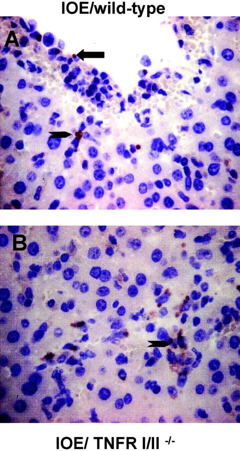
Intraperitoneal (i.p.) infection with a high dose of a highly virulent Ehrlichia strain (IOE) results in a toxic shock-like syndrome characterized by severe liver injury and systemic overproduction of tumor necrosis factor alpha (TNF-alpha) by CD8+ T cells. We examined the role of TNF-alpha and TNF receptors in high-dose-IOE-induced shock/liver injury. TNF receptor (TNFR) I/II-/- mice lacking both the p55 and p75 receptors for this cytokine were more resistant to IOE-induced liver injury than their wild-type background controls. TNFR I/II-/- mice survived longer, dying between 15 and 18 days, with evidence of mild liver necrosis/apoptosis. In contrast, wild-type mice were not rescued from the lethal effect of IOE by TNF-alpha neutralization. TNF-alpha-depleted mice developed severe liver injury and succumbed to disease between days 9 and 11 postinfection, similar to sham-treated, infected wild-type mice. Although IFN-gamma production in the spleens of IOE-infected TNFR I/II-/- and TNF-alpha-depleted mice was higher than that detected in wild-type controls, these mice had higher bacterial burdens than infected controls. Following high-dose IOE challenge, TNFR I/II-/- and TNF-alpha-depleted mice have an early increase in IL-10 levels in sera and spleens, which was produced mainly by adherent spleen cells. In contrast, a late burst of interleukin-10 (IL-10) was observed in control mice. Nonadherent spleen cells were the major source of IL-10 in IOE-infected wild-type mice. We conclude that TNFR I/II and TNF-alpha participate in Ehrlichia-induced shock and host defense by regulating liver injury and controlling ehrlichial burden. Our data suggest that fatal ehrlichiosis could be a multistep process, where TNF-alpha is not solely responsible for mortality.


Figures

FIG. 1.
FIG. 1.
Increased resistance of TNFR I/II−/− mice to IOE-induced toxic shock. (A) Survival rates of various mouse groups following challenge with a high dose of highly virulent IOE or mildly virulent E. muris. Six- to 8-week-old C57BL/6 control (n = 12) mice and mice treated with anti-TNF-α mAb (n = 12) were infected i.p. with a high dose of IOE or E. muris 1 day after the initiation of TNF-α neutralization. TNFR I/II−/− mice (n = 15) were infected i.p. with the same dose of IOE. Mice were monitored daily for disease manifestations and death. The data shown represent one of three independent experiments. WT, wild type. (B and C) Higher bacterial burdens in different organs in anti-TNF-α Ab-treated mice (B) and TNFR I/II−/− mice (C) than wild-type controls on day 8 following i.p. infection with IOE, as determined by real-time PCR. Significance was determined by Student's two-tailed t test (symbols above bars indicate P < 0.001 for IOE-infected anti-TNF-α Ab-treated mice versus IOE-infected wild-type mice [B] or for IOE-infected TNFR I/II−/− KO mice versus IOE-infected wild-type mice [C]). Data represent the averages and standard deviations of triplicate amplifications with three mice per group. Similar results were observed in three independent experiments.
FIG. 1.
FIG. 1.
Increased resistance of TNFR I/II−/− mice to IOE-induced toxic shock. (A) Survival rates of various mouse groups following challenge with a high dose of highly virulent IOE or mildly virulent E. muris. Six- to 8-week-old C57BL/6 control (n = 12) mice and mice treated with anti-TNF-α mAb (n = 12) were infected i.p. with a high dose of IOE or E. muris 1 day after the initiation of TNF-α neutralization. TNFR I/II−/− mice (n = 15) were infected i.p. with the same dose of IOE. Mice were monitored daily for disease manifestations and death. The data shown represent one of three independent experiments. WT, wild type. (B and C) Higher bacterial burdens in different organs in anti-TNF-α Ab-treated mice (B) and TNFR I/II−/− mice (C) than wild-type controls on day 8 following i.p. infection with IOE, as determined by real-time PCR. Significance was determined by Student's two-tailed t test (symbols above bars indicate P < 0.001 for IOE-infected anti-TNF-α Ab-treated mice versus IOE-infected wild-type mice [B] or for IOE-infected TNFR I/II−/− KO mice versus IOE-infected wild-type mice [C]). Data represent the averages and standard deviations of triplicate amplifications with three mice per group. Similar results were observed in three independent experiments.
FIG. 2.
FIG. 2.
Immunohistochemical (IHC) detection of IOE in the livers of mice infected with a high dose of IOE. (A) Isotype control wild-type mice; (B) TNFR I/II−/− mice. IHC detection in livers from TNFR I/II−/− mice demonstrated numerous morulae in sinusoidal and blood vessel lining cells (Kupffer cells and endothelial cells) as well as in hepatocytes (arrowhead). In contrast, IHC detection in livers of IOE-infected wild-type control mice demonstrated fewer morulae, which were detected mainly in sinusoidal-lining cells, including endothelial and Kupffer cells (arrow). A horseradish peroxidase (diaminobenzidine)/hematoxylin stain was used. Original magnification, ×400.
FIG. 3.
FIG. 3.
Markedly decreased liver injury/apoptosis in TNFR I/II−/− mice. (A) Wild-type mice treated with isotype control; (B) anti-TNF-α-treated mice; (C) TNFR I/II−/− mice. All mice were infected i.p. with a high dose of IOE, and livers were collected at 8 days postinfection and stained with H&E (A to D). Extensive necrosis was observed in wild-type and anti-TNF-α-treated mice (arrows), while few apoptotic cells were detected in TNFR I/II−/− mice (arrowhead). H&E staining is shown. Original magnifications, ×200 (A and D), ×100 (C), and ×400 (B). (D) A high-power view of the livers of TNFR I/II−/− treated mice showed steatosis with middle- (arrow) and large-sized-droplet fatty change. Anti-TNF-α-treated mice had similar fatty change (not shown). (E) TUNEL assays using horseradish peroxidase (diaminobenzidine)/azure A stain of livers of wild-type mice showed extensive apoptosis of hepatocytes (arrow) and cells lining hepatic sinusoids such as Kupffer cells and endothelial cells (arrow). Original magnification, ×400.
FIG. 3.
FIG. 3.
Markedly decreased liver injury/apoptosis in TNFR I/II−/− mice. (A) Wild-type mice treated with isotype control; (B) anti-TNF-α-treated mice; (C) TNFR I/II−/− mice. All mice were infected i.p. with a high dose of IOE, and livers were collected at 8 days postinfection and stained with H&E (A to D). Extensive necrosis was observed in wild-type and anti-TNF-α-treated mice (arrows), while few apoptotic cells were detected in TNFR I/II−/− mice (arrowhead). H&E staining is shown. Original magnifications, ×200 (A and D), ×100 (C), and ×400 (B). (D) A high-power view of the livers of TNFR I/II−/− treated mice showed steatosis with middle- (arrow) and large-sized-droplet fatty change. Anti-TNF-α-treated mice had similar fatty change (not shown). (E) TUNEL assays using horseradish peroxidase (diaminobenzidine)/azure A stain of livers of wild-type mice showed extensive apoptosis of hepatocytes (arrow) and cells lining hepatic sinusoids such as Kupffer cells and endothelial cells (arrow). Original magnification, ×400.
FIG. 4.
FIG. 4.
Serum levels of TNF-α and IL-12 and frequencies of Ag-specific IFN-γ- and IL-4-producing cells in the spleens of TNF-α-depleted, TNFR I/II−/−, and wild-type mice treated with isotype control following challenge with either IOE or E. muris. Mice were infected i.p. with either a high dose of IOE or E. muris (A to C). Sera were collected at 7 days for TNF-α (A) and at 3 days [for IL-12 (p40) and Il-12 (p70) (B and C, respectively)] after challenge, and serum levels of these cytokines were determined by ELISA. The serum level of TNF-α was lower in IOE-infected anti-TNF-α-treated mice versus infected wild-type controls (solid star, P < 0.001), but serum TNF-α was higher in TNFR I/II−/− KO versus wild-type controls (solid diamond, P < 0.005). Serum levels of IL-12 (p40) and IL-12 (p70) were slightly higher in TNFR I/II−/− KO mice versus wild-type controls [open star, P > 0.05 for both IL-12 (p40) and IL-12 (p70)]. Serum levels of IL-12 (p40) and IL-12 (p70) were significantly lower in TNFR I/II−/− KO mice versus E. muris-infected control mice (four diamonds, P < 0.001). Before Ehrlichia challenge, the above cytokines were virtually undetectable in the sera. (D) Spleens were collected at day 7 postinfection, and splenocytes were prepared thereafter and stimulated in vitro with the relevant antigen. The frequencies of IFN-γ and IL-4 were determined by ELISPOT assay. Data represent the averages and standard deviations of four mice per group. Similar results were observed in three independent experiments, with a total of 12 mice/group.
FIG. 4.
FIG. 4.
Serum levels of TNF-α and IL-12 and frequencies of Ag-specific IFN-γ- and IL-4-producing cells in the spleens of TNF-α-depleted, TNFR I/II−/−, and wild-type mice treated with isotype control following challenge with either IOE or E. muris. Mice were infected i.p. with either a high dose of IOE or E. muris (A to C). Sera were collected at 7 days for TNF-α (A) and at 3 days [for IL-12 (p40) and Il-12 (p70) (B and C, respectively)] after challenge, and serum levels of these cytokines were determined by ELISA. The serum level of TNF-α was lower in IOE-infected anti-TNF-α-treated mice versus infected wild-type controls (solid star, P < 0.001), but serum TNF-α was higher in TNFR I/II−/− KO versus wild-type controls (solid diamond, P < 0.005). Serum levels of IL-12 (p40) and IL-12 (p70) were slightly higher in TNFR I/II−/− KO mice versus wild-type controls [open star, P > 0.05 for both IL-12 (p40) and IL-12 (p70)]. Serum levels of IL-12 (p40) and IL-12 (p70) were significantly lower in TNFR I/II−/− KO mice versus E. muris-infected control mice (four diamonds, P < 0.001). Before Ehrlichia challenge, the above cytokines were virtually undetectable in the sera. (D) Spleens were collected at day 7 postinfection, and splenocytes were prepared thereafter and stimulated in vitro with the relevant antigen. The frequencies of IFN-γ and IL-4 were determined by ELISPOT assay. Data represent the averages and standard deviations of four mice per group. Similar results were observed in three independent experiments, with a total of 12 mice/group.
FIG. 5.
FIG. 5.
Levels of IL-10 in the sera and spleen and frequencies of IL-10-producing spleen cells in TNF-α-depleted, TNFR I/II−/−, and wild-type mice following lethal or nonlethal challenge with IOE or E. muris, respectively. Mice were infected i.p. with either a high dose of IOE or E. muris. (A) Sera were collected at 3, 8, and 9 days after challenge, and serum levels of IL-10 were determined by ELISA. Before Ehrlichia challenge, the above cytokines were virtually undetectable in the sera. (B) Spleens were collected at day 8 postinfection, and splenocytes were prepared thereafter and stimulated in vitro with the relevant antigen. The frequencies of Ag-specific IL-10-producing cells were determined by ELISPOT assay. The numbers of Ag-specific IL-10-producing cells were comparable among all IOE-infected mice in different groups (P > 0.05) but were significantly higher than that detected in E. muris-infected mice (P < 0.0005). Data represent the averages and standard deviations of four mice per group. Similar results were observed in three independent experiments, with a total of nine mice/group.
FIG. 6.
FIG. 6.
IL-10 production by total unseparated, nonadherent and adherent spleen cells from wild-type, TNFR I/II−/−, and anti-TNF-α-treated mice after in vitro stimulation with IOE. All mice were infected with IOE, and spleens were recovered on day 8 postinfection. Supernatant was collected at 48 h, and the IL-10 level was measured by ELISA. In wild-type mice, IL-10 was secreted mainly by adherent cells in an antigen-dependent manner, while in TNFR I/II−/− and anti-TNF-α-treated mice, IL-10 was mainly produced by adherent cells. Solid star, four diamonds, P < 0.005 for IL-10 produced by adherent or nonadherent cells from IOE-infected wild-type versus TNFR I/II−/− KO mice; solid diamond, open star, P < 0.001 for IL-10 produced by adherent or nonadherent cells from IOE-infected wild-type versus anti-TNF-α-depleted mice. Bars represent means ± standard deviations of three independent observations conducted in duplicate.

Similar articles

Cited by

References

    1. Bean, A. G., D. R. Roach, H. Briscoe, M. P. France, H. Korner, J. D. Sedgwick, and W. J. Britton. 1999. Structural deficiencies in granuloma formation in TNF gene-targeted mice underlie the heightened susceptibility to aerosol Mycobacterium tuberculosis infection, which is not compensated for by lymphotoxin. J. Immunol. 162:3504-3511. - PubMed
    1. Bitsaktsis, C., J. Huntington, and G. Winslow. 2004. Production of IFN-gamma by CD4 T cells is essential for resolving ehrlichia infection. J. Immunol. 172:6894-6901. - PubMed
    1. Bogdan, C., M. Rollinghoff, and A. Diefenbach. 2002. The role of nitric oxide in innate immunity. Immunol. Rev. 173:17-26. - PubMed
    1. Chadban, S. J., G. H. Tesch, R. Foti, H. Y. Lan, R. C. Atkins, and D. J. Nikolic-Paterson. 1998. Interleukin-10 differentially modulates MHC class II expression by mesangial cells and macrophages in vitro and in vivo. Immunology 94:72-78. - PMC - PubMed
    1. Chan, K. F., M. R. Siegel, and J. M. Lenardo. 2000. Signaling by the TNF receptor superfamily and T cell homeostasis. Immunity 13:419-422. - PubMed

Publication types

Substances

LinkOut - more resources