. 2006 Mar;74(3):1924-32.
doi: 10.1128/IAI.74.3.1924-1932.2006.

Amelioration of influenza-induced pathology in mice by coinfection with Trichinella spiralis

Affiliations

Amelioration of influenza-induced pathology in mice by coinfection with Trichinella spiralis

Rebecca C Furze et al. Infect Immun. 2006 Mar.

Abstract

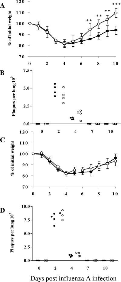
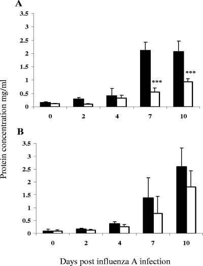
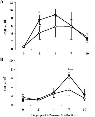
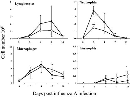
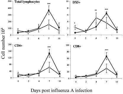
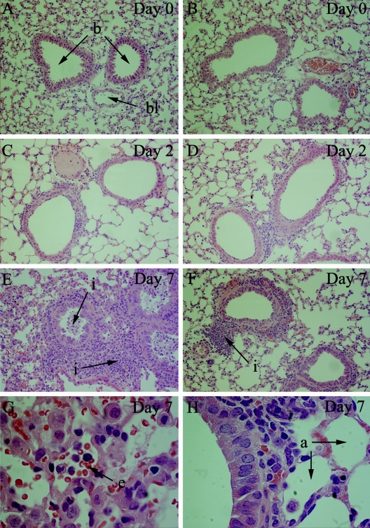
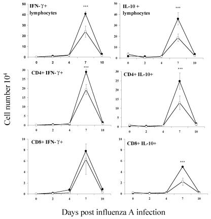
Illness due to respiratory virus infection is often induced by excessive infiltration of cells into pulmonary tissues, leading to airway occlusion. We show here that infection with Trichinella spiralis results in lower levels of tumor necrosis factor in bronchoalveolar lavage fluid and inhibits cellular recruitment into the airways of mice coinfected with influenza A virus. Infiltration of neutrophils and CD4+ and CD8+ lymphocytes was reduced, resulting in animals gaining weight more rapidly following the initial phase of infection. Influenza resulted in a generalized increase in vascular permeability in pulmonary tissues, and this was suppressed by parasite infection, although the effects were restricted to the early phase of trichinosis. Moreover, the number of cells producing interleukin-10 (IL-10), and the local levels of this cytokine, were reduced, suggesting that amelioration of pulmonary pathology by parasite infection occurs independently of IL-10 production.


Figures

FIG. 1.
FIG. 1.
Coinfection with enteric-stage T. spiralis infection enhances recovery from influenza infection but has no effect on viral clearance. Mice were infected orally with T. spiralis and at two later distinct time points inoculated intranasally with 50 HA units of influenza A virus (X31). Weight loss was monitored daily and expressed as a percentage of the weight prior to influenza infection (influenza administered at day 7 [A] and at day 60 [C] of parasite infection). Viral load per lung was assessed by plaque assay (influenza administered at day 7 [B] and at day 60 [D] of parasite infection). Points in panels B and D represent viral titers from individual mice. In all panels, data for mice infected with influenza virus alone are shown as filled circles and data for coinfected animals shown as open circles; data represent mean values ± 1 SD. Significance values are shown relative to mice infected solely with influenza. **, P ≤ 0.01; ***, P ≤ 0.001.
FIG. 2.
FIG. 2.
Coinfection results in lower albumin concentrations in BAL fluid. Mice were infected orally with T. spiralis and 7 days (A) or 60 days (B) later inoculated intranasally with influenza A virus. The albumin concentration in lavage fluid was determined at different time points after viral infection. Filled bars indicate data for mice infected with influenza alone and open bars represent data for coinfected animals. Mean values ± 1 SD are shown, and significant differences relative to mice infected solely with influenza are highlighted. ***, P ≤ 0.001.
FIG. 3.
FIG. 3.
Cellular infiltration into the lungs is suppressed by T. spiralis. Mice were inoculated with influenza virus 7 days following infection with T. spiralis. Total cells recovered from the BAL fluid (A) and lungs (B) were enumerated over the course of viral infection. Values represent means ± 1 SD, and significant differences to mice infected with influenza virus alone are shown. Filled circles, influenza infection alone; open circles, coinfected animals. *, P ≤ 0.05; ***, P ≤ 0.001.
FIG. 4.
FIG. 4.
Parasite infection inhibits the early influx of neutrophils. Cells recovered from BAL fluid at different times after influenza infection were stained with hematoxylin and eosin following cytospin preparation. Neutrophils, eosinophils, lymphocytes, and macrophages were enumerated. Data are presented from mice infected solely with influenza virus and from those infected 7 days earlier with T. spiralis. The values shown are means ± 1 SD, and significance values for coinfected mice (open circles) are expressed in comparison to mice infected with influenza virus alone (filled circles). ***, P ≤ 0.001.
FIG. 5.
FIG. 5.
Reduction and altered kinetics of lymphocyte infiltration. Mice were infected with T. spiralis and with influenza A virus 7 days later, or influenza A virus alone, as described. Single-cell suspensions from the lungs were analyzed by flow cytometry, and total numbers of lymphocytes and CD4+, CD8+, and DX5+ cells were determined. Numbers are expressed as means ± 1 SD, and significant differences between singly infected (filled circles) and coinfected (open circles) mice are shown. **, P ≤ 0.01; ***, P ≤ 0.001.
FIG. 6.
FIG. 6.
Amelioration of cellular infiltration and airway occlusion. Lungs were fixed, sectioned, and stained with hematoxylin and eosin. (A, C, E, and G) Tissue from mice infected with influenza virus alone is shown; (B, D, F, and H) tissue from mice preinfected with T. spiralis is shown. Representative sections from each group are shown at day 0 (A and B), day 2 (C and D), and day 7 (E and F) after influenza infection at 40× magnification. Sections of lungs at day 7 p.i. are also shown at 100× magnification (G and H). Bronchioles (b), alveoli (a), and blood vessels (bl) are shown. Infiltrating cells (i) are indicated, as are erythrocytes (e) in the alveolar spaces.
FIG. 7.
FIG. 7.
Infiltration of both IFN-γ- and IL-10-secreting lymphocytes is suppressed by parasite infection. Samples were analyzed by flow cytometry and the numbers of IFN-γ- and IL-10-positive lymphocytes in the lung determined by intracellular cytokine staining. Numbers are expressed as means ± 1 SD, and significant differences between singly infected (filled circles) and coinfected (open circles) mice are shown. ***, P ≤ 0.001.
FIG. 8.
FIG. 8.
Prior infection with T. spiralis suppresses IL-10 and TNF-α in BAL fluid. Concentrations of cytokines were determined by enzyme-linked immunosorbent assay. The values shown represent the means ± 1 SD, and significant differences between singly infected (filled bars) and coinfected (open bars) mice are shown. ***, P ≤ 0.001.
FIG. 9.
FIG. 9.
Reduced numbers of T lymphocytes in the lungs are not accounted for by enhanced apoptosis or suppression of proliferation. Mice were infected orally with T. spiralis and 7 days later with influenza A virus as described. Single-cell suspensions from the MdLN were analyzed by flow cytometry and apoptotic cells defined as annexin+ and To-pro-3. The proportions of apoptotic CD4+ (A) and CD8+ (B) lymphocytes were determined. Cellular proliferation was assessed by incorporation of BrdU and flow cytometry (C). No significant differences were observed between singly infected (filled bars) or coinfected (open bars) animals in any case.

Similar articles

Cited by

References

    1. Actor, J. K., M. Shirai, M. C. Kullberg, R. M. Buller, A. Sher, and J. A. Berzofsky. 1993. Helminth infection results in decreased virus-specific CD8+ cytotoxic T-cell and Th1 cytokine responses as well as delayed virus clearance. Proc. Natl. Acad. Sci. USA 90:948-952. - PMC - PubMed
    1. Bany, J., M. K. Janiak, and W. Budzynski. 1992. Activity of natural killer (NK) cells in the course of experimental trichinellosis in mice. Wiad. Parazytol. 38:117-126. - PubMed
    1. Beiting, D. P., S. K. Bliss, D. H. Schlafer, V. L. Roberts, and J. A. Appleton. 2004. Interleukin-10 limits local and body cavity inflammation during infection with muscle-stage Trichinella spiralis. Infect. Immun. 72:3129-3137. - PMC - PubMed
    1. Biron, C. A., and L. Brossay. 2001. NK cells and NKT cells in innate defense against viral infections. Curr. Opin. Immunol. 13:458-464. - PubMed
    1. Bliss, S. K., A. Alcaraz, and J. A. Appleton. 2003. IL-10 prevents liver necrosis during murine infection with Trichinella spiralis. J. Immunol. 171:3142-3147. - PubMed

Publication types