Comparative Study
. 2006 Mar;243(3):389-98.
doi: 10.1097/01.sla.0000201541.68577.6a.

A rapid, fully automated, molecular-based assay accurately analyzes sentinel lymph nodes for the presence of metastatic breast cancer

Affiliations
Comparative Study

A rapid, fully automated, molecular-based assay accurately analyzes sentinel lymph nodes for the presence of metastatic breast cancer

Steven J Hughes et al. Ann Surg. 2006 Mar.

Abstract

Objective: To develop a fully automated, rapid, molecular-based assay that accurately and objectively evaluates sentinel lymph nodes (SLN) from breast cancer patients.

Summary background data: Intraoperative analysis for the presence of metastatic cancer in SLNs from breast cancer patients lacks sensitivity. Even with immunohistochemical staining (IHC) and time-consuming review, alarming discordance in the interpretation of SLN has been observed.

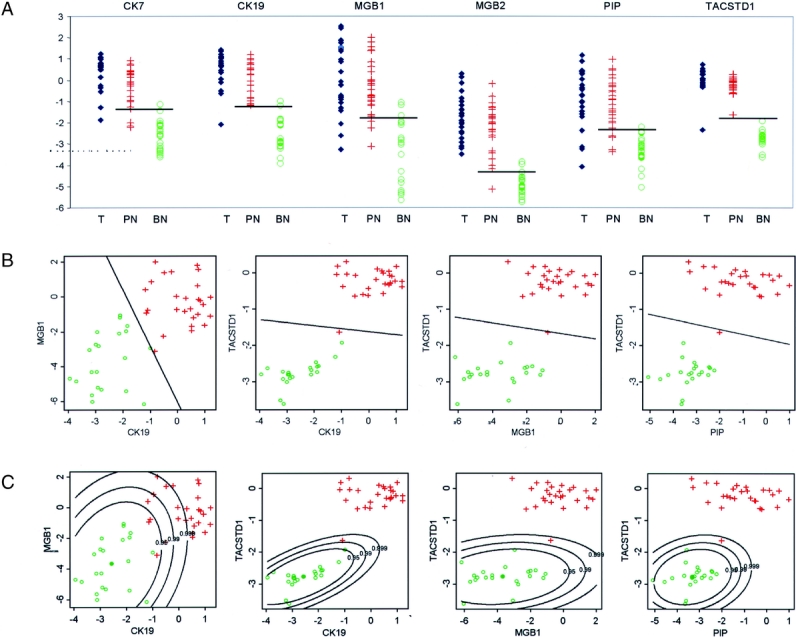
Method: A total of 43 potential markers were evaluated for the ability to accurately characterize lymph node specimens from breast cancer patients as compared with complete histologic analysis including IHC. Selected markers then underwent external validation on 90 independent SLN specimens using rapid, multiplex quantitative reverse transcription-polymerase chain reaction (QRT-PCR) assays. Finally, 18 SLNs were analyzed using a completely automated RNA isolation, reverse transcription, and quantitative PCR instrument (GeneXpert).

Results: : Following analysis of potential markers, promising markers were evaluated to establish relative level of expression cutoff values that maximized classification accuracy. A validation set of 90 SLNs from breast cancer patients was prospectively characterized using 4 markers individually or in combinations, and the results compared with histologic analysis. A 2-marker assay was found to be 97.8% accurate (94% sensitive, 100% specific) compared with histologic analysis. The fully automated GeneXpert instrument produced comparable and reproducible results in less than 35 minutes.

Conclusions: A rapid, fully automated QRT-PCR assay definitively characterizes breast cancer SLN with accuracy equal to conventional pathology. This approach is superior to intraoperative SLN analysis and can provide standardized, objective results to assist in pathologic diagnosis.


Figures

None
FIGURE 1. A, Data obtained from the secondary screening set of lymph nodes on individual gene expression observed in primary tumors, benign and positive lymph nodes. The horizontal line indicates the most accurate cutoff value for classifying the lymph node as positive or benign (T, tumor; PN, positive node; BN, benign node). B, Secondary screening set data on gene expression for potential 2-marker combinations using a linear discriminator decision rule. The line equalizes the predicted probability that a lymph node is positive or benign. (green o = benign lymph node; red + = histologically positive lymph node). C, Secondary screening set data on gene expression for potential 2-marker combinations applying equal probability contour statistical analysis. Equal probability curves were generated around the mean expression value observed for the 2 markers in benign lymph nodes. This demonstrates that while the marker combination of CK19 and MGB1 accurately characterizes the lymph nodes (Table 2), the wide distribution of expression observed in benign nodes diminishes their ability as accurate predictors. By this method of analysis, the marker combination of TACSTD1 and PIP more confidently characterizes the lymph nodes.
None
FIGURE 2. A, Data obtained from the validation set of SLN on individual gene expression observed in negative and positive lymph nodes (green o = histologically negative lymph node; red + = histologically positive lymph node). The horizontal line indicates the decision rule calculated from data obtained from the secondary screening set. The line equalizes the predicted probability that a lymph node is positive or negative. B, Validation set data on gene expression for 2-marker combinations using linear discriminator decision rule for all potential marker pairs. Classification characteristics of the marker combinations are reported in Table 3. C, Validation set data analyzed using the equal probability contours generated from the secondary screening set data. The relative levels of expression observed for the positive lymph nodes were beyond the 0.999 probability contours for 3 2-marker combinations. Some of the positive nodes are positive for only one marker (red crosses located in the left upper or right lower quadrants), demonstrating how a 2-marker assay improves sensitivity while maintaining high specificity.
None
FIGURE 3. Results of a fully automated, 2-marker QRT-PCR analysis of lymph nodes. By either linear decision rule analysis (left panel) or equal probability contour analysis (right panel), the assay accurately characterized all 18 lymph nodes (9 negative, 9 positive) evaluated (green o = histologically negative lymph node; red + = histologically positive lymph node).
None
FIGURE 4. Proposed method of lymph node preparation that facilitates thorough histologic analysis and isolation of high-quality RNA. The lymph node is cut into 5 sections. Three sections are processed into paraffin for subsequent histologic analysis. Two sections are embedded in OCT and cryostat sections obtained for histologic analysis and RNA isolation. The remaining uncut tissue may be stored frozen or paraffin-embedded for future analysis.

References

    1. Fisher B, Bauer M, Wickerham DL, et al. Relation of number of positive axillary nodes to the prognosis of patients with primary breast cancer: an NSABP update. Cancer. 1983;52:1551–1557. - PubMed
    1. Fisher ER, Sass R, Fisher B. Pathologic findings from the National Surgical Adjuvant Project for Breast Cancers (protocol no. 4): X. Discriminants for tenth year treatment failure. Cancer. 1984;53:712–723. - PubMed
    1. Fisher ER, Costantino J, Fisher B, et al. Pathologic findings from the National Surgical Adjuvant Breast Project (Protocol 4): Discriminants for 15-year survival. National Surgical Adjuvant Breast and Bowel Project Investigators. Cancer. 1993;71:2141–2150. - PubMed
    1. Siziopikou KP, Schnitt SJ, Connolly JL, et al. Detection and significance of occult axillary metastatic disease in breast cancer patients. Breast J. 1999;5:221–229. - PubMed
    1. Turner RR, Ollila DW, Krasne DL, et al. Histopathologic validation of the sentinel lymph node hypothesis for breast carcinoma. Ann Surg. 1997;226:271–276. - PMC - PubMed

Publication types