. 2006 Apr;12(4):631-43.
doi: 10.1261/rna.2226106. Epub 2006 Feb 22.

AtCyp59 is a multidomain cyclophilin from Arabidopsis thaliana that interacts with SR proteins and the C-terminal domain of the RNA polymerase II

Affiliations

AtCyp59 is a multidomain cyclophilin from Arabidopsis thaliana that interacts with SR proteins and the C-terminal domain of the RNA polymerase II

Monika Gullerova et al. RNA. 2006 Apr.

Abstract

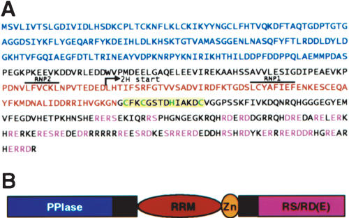
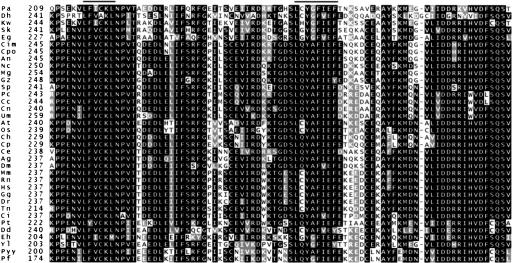
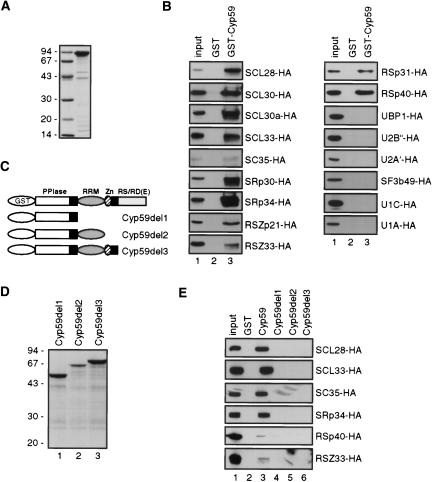
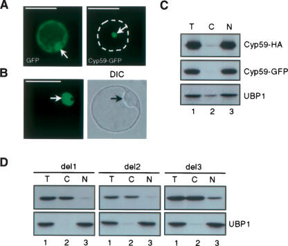
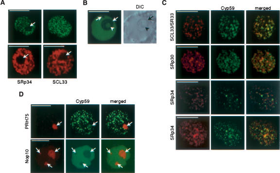
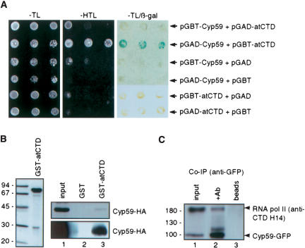
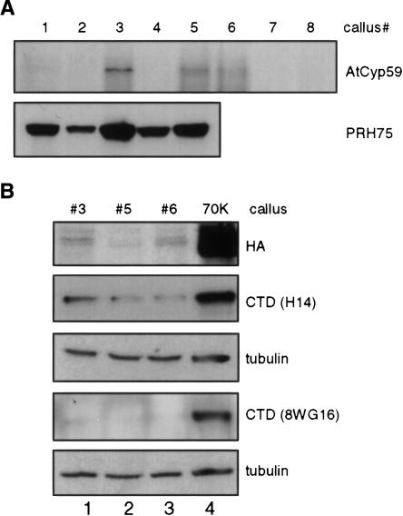
AtCyp59 and its orthologs from different organisms belong to a family of modular proteins consisting of a peptidyl-prolyl cis-trans isomerase (PPIase) domain, followed by an RNA recognition motif (RRM), and a C-terminal domain enriched in charged amino acids. AtCyp59 was identified in a yeast two-hybrid screen as an interacting partner of the Arabidopsis SR protein SCL33/SR33. The interaction with SCL33/SR33 and with a majority of Arabidopsis SR proteins was confirmed by in vitro pull-down assays. Consistent with these interactions, AtCyp59 localizes to the cell nucleus, but it does not significantly colocalize with SR proteins in nuclear speckles. Rather, it shows a punctuate localization pattern resembling transcription sites. Indeed, by using yeast two-hybrid, in vitro pull-down, and immunoprecipitation assays, we found that AtCyp59 interacts with the C-terminal domain (CTD) of the largest subunit of RNA polymerase II. Ectopic expression of the tagged protein in Arabidopsis cell suspension resulted in highly reduced growth that is most probably due to reduced phosphorylation of the CTD. Together our data suggest a possible function of AtCyp59 in activities connecting transcription and pre-mRNA processing. We discuss our data in the context of a dynamic interplay between transcription and pre-mRNA processing.


Figures

FIGURE 1.
FIGURE 1.
Sequence analysis of AtCyp59. (A) Protein sequence of Arabidopsis AtCyp59. The peptidyl-prolyl cistrans isomerase (PPIase) domain is printed in blue letters, and the RNA recognition motif, (RRM) in red letters with RNP1 and RNP2 overlined. The CCHC zinc knuckle is printed on yellow background with CCHC in green letters. The RS, RD, and RE dipeptides are printed in pink letters. Position of the start of two-hybrid clone isolated with SCL33/SR33 is indicated by an arrow. (B) Schematic representation of AtCyp59 modular structure.
FIGURE 2.
FIGURE 2.
Sequence alignment of AtCyp59 RRM with corresponding domains from different organisms. Sequences were aligned by using ClustalW and shaded on BoxShade server. Amino acids identical or similar in 50% of sequences are shaded on black or on gray background, respectively. Order of the sequences is as appeared in ClustalW output. The RNP1 and RNP2 motifs of RRM are overlined. Hs, Homo sapiens; Mm, Mus musculus; Rn, Rattus norvegicus; Ci, Cionia intestinalis; Dr, Danio rerio; Gg, Gallus gallus; Tn, Tetraodon nigrovirides; Ce, Caenorhabditis elegans; Dm, Drosophila melanogaster; Ag, Aenopheles gambiae; At, Arabidopsis thaliana; Os, Oryza sativa; Ch, Cryptosporidium hominis; Cp, Cryptosporidium parvum; Pf, Plasmodium falciparum; Pyy, Plasmodium yoelii yoelii; Dd, Dictyostelium discoideum; Eh, Entamoeba histolytica; Pt, Paramecium tetraurelia; Sp, SchizoSaccharomyces pombe; Nc, Neurospora crassa; Mg, Magnaportae grisea; Gz, Gibberella zeae; An, Aspergillus nidulans; Cpo, Coccidioides posadasii; Cim, Coccidioides immitis; Eg, Eremothecium gossypii; Dh, Debaryomyces hansenii; Kw, Kluyveromyces waltii; Sk, Saccharomyces kluyveri; Yl, Yarrowia lipolytica; Cc, Cryptococcus cinereea okayama; Cn, Cryptococcus neoformans; Pc, Phanerochaete chrysosporium; Um, Ustilago maydis; Pa, Pichia angusta.
FIGURE 3.
FIGURE 3.
Interaction of AtCyp59 with SR proteins. (A) Coomassie blue–stained gel of purified recombinant GST-AtCyp59. Molecular mass standards in kilodaltons are indicated on the left. (B) AtCyp59 interacts with SR proteins in vitro. Whole cell extracts from protoplasts transiently expressing HA-tagged SR proteins were incubated with glutathione Sepharose beads coated with GST-AtCyp59. After washing, proteins retained on the beads were analyzed by SDS-PAGE and Western blotting with rat anti-HA monoclonal antibody. (Lanes 1) 1/10 of the input extract used for pull-downs in lanes 2 and 3; (lanes 2) pull-downs with beads coated with GST alone; (lanes 3) pull-downs with beads coated GST-AtCyp59. (C) Schematic representation of AtCyp59 deletion mutants fused to GST. (D) Coomassie blue–stained gel of purified recombinant GST-AtCyp59 deletion mutants. Molecular mass standards in kilodaltons are indicated on the left. (E) Interaction of AtCyp59 deletion mutants with SR proteins. Pull-down experiment performed with GST-AtCyp59 deletion mutants and one member of each Arabidopsis SR protein subfamilies. (Lane 1) 1/10 of the input extract used for pull-downs in lanes 26; (lane 2) pull-downs with beads alone; (lane 3) pull-downs with beads coated GST-AtCyp59; (lane 4) pull-downs with beads coated GST-AtCyp59D1; (lane 5) pull-downs with beads coated GST-AtCyp59D2; (lane 6) pull-downs with beads coated GST-AtCyp59D3.
FIGURE 4.
FIGURE 4.
Determination of RNA binding specificity of AtCyp59. Nucleotide binding specificity was measured by the UV cross-linking/homoribopolymer competition assay using 32P-labeled Syn7 RNA and recombinant GST-AtCyp59 protein. Different indicated polymers were added at 10-fold (lanes 2,6,10,14), 50-fold (lanes 3,7,11,15), 200-fold (lanes 4,8,12,16), and 400-fold (lanes 5,9,13,17) excess over Syn7 RNA (calculated in moles of nucleotides). Minus sign (–) (lanes 1,18), cross-linking without any competitor added.
FIGURE 5.
FIGURE 5.
Cellular localization of AtCyp59. (A) Localization of AtCyp59-GFP fusion protein and GFP alone in transiently transformed tobacco protoplasts. Dashed line delineates the shape of protoplasts. Arrows point to nuclei. (B) Localization of AtCyp59-GFP fusion protein in transiently transformed Arabidopsis protoplasts. Single confocal image with corresponding differential interference contrast (DIC) image of whole cell is shown. Arrows point to nuclei. Protoplasts were analyzed 24 h after transformation by using Zeiss Axiovert epifluorescence microscope (A) or Leica TCS confocal microscope (B). Bars, 50 μm (A,B). (C) Cellular localization of AtCyp59 studied by cellular fractionation of Arabidopsis protoplasts transiently expressing AtCyp59-GFP or AtCyp59-HA. Western blots were analyzed with monoclonal antibodies against GFP or HA tags. The distribution of endogenous nuclear UBP1 protein was used to control quality of the fractionation procedure. (Lane 1) Total protein extracts (T), (lane 2) cytoplasmic fraction (C), (lane 3) nuclear fraction (N). (D) Localization of AtCyp59-HA deletions in transiently transformed Arabidopsis protoplasts as determined by cellular fractionation. Details are as in C.
FIGURE 6.
FIGURE 6.
Subnuclear localization of AtCyp59. (A) Cofocal images of nuclei from tobacco cells expressing AtCyp59-GFP (single confocal section, left; maximum intensity projection of all sections of the same nucleus, right) and two SR proteins, SRp34 and SCL33 (two lower panels; shown are only single confocal sections). Note that AtCyp59-GFP localizes into punctuate pattern, which is different from speckled pattern of SR proteins. Arrows point to nucleoli. Bars, 8 μm. (B) Single confocal section of Arabidopsis nucleus expressing AtCyp59-GFP (left) with corresponding DIC image (right). Arrows point to nucleoli and arrowhead to nucleolar cavity. Bar, 8 μm. (C) Colocalization studies of AtCyp59 with Arabidopsis SR proteins. Tobacco protoplasts were transiently cotransformed with plasmids expressing AtCyp59-GFP and indicated SR proteins fused to RFP. Maximum intensity projections of cotransformed nuclei are shown. For SRp34–AtCyp59 combination also a single confocal section is shown (second row from bottom). Merged images show superimposition of GFP and RFP signals. Bars, 7 μm. (D) Colocalization studies of AtCyp59 with markers for nucleoli [PRH75-RFP (tobacco protoplasts) and Nop10-RFP (Arabidopsis protoplasts)]. Only single confocal sections are shown. Arrows point to nucleoli. Bars, 5 μm.
FIGURE 7.
FIGURE 7.
AtCyp59 interacts with the RNA polymerase II CTD. (A) Interaction between AtCyp59 and CTD in a yeast two-hybrid assay. Indicated combinations of plasmids were cotrans-formed into yeast reporter strain, and the interaction of AtCyp59 with the CTD was assessed by growth on plates lacking histidine (–HTL) or by analysis of the activation of the second reporter gene, β-galactosidase (–HTL/β-gal). (B) In vitro interaction between AtCyp59 and CTD studied by pull-down assay. Arabidopsis CTD was overexpressed as a GST fusion in E. coli and purified (left panel). Pull-downs with AtCyp59-HA expressed in Arabidopsis protoplasts (upper right panel) or in E. coli (lower right panel). (Lanes 1) 1/10 of the input protein extracts used in pull-downs in lanes 2 and 3. (Lanes 2) pull-down with GST alone. (Lanes 3) pull-down with GST-AtCyp59. (C) In vivo interaction of AtCyp59 and CTD determined by coimmunoprecipitation. Protein extract from protoplasts transiently expressing AtCyp59-GFP were immunoprecipitated with anti-GFP antibody. Blot was probed with anti-GFP and anti-CTD (H14) antibodies simultaneously. (Lane 1) 1/ 10 of the protein extract used in IPs in lanes 2 and 3; (lane 2) immunoprecipitation with anti-GFP antibody; (lane 3) protein extract incubated with Protein A beads only.
FIGURE 8.
FIGURE 8.
Stable expression of AtCyp59 in Arabidopsis cells results in cell growth arrest and decrease in CTD phosphorylation. (A) Western blot analysis of transgenic, kanamycin resistant Arabidopsis calli expressing TAP tagged AtCyp59 (upper panel). For comparison transgenic calli expressing TAP-tagged PRH75 (lower panel) were analyzed in parallel. Both proteins were detected with anti-HA antibody. (B) Analysis of the phosphorylation status of the RNA pol II CTD in Arabidopsis calli expressing TAP-tagged AtCyp59. (Top panel) Analysis of the expression of AtCyp59 (lanes 13). For comparison, protein extract from callus expressing Arabidopsis U1 snRNP-specific protein 70K was loaded as a control (lane 4). The same protein extracts were analyzed with antibodies against the CTD (H14, second panel, and 8WG16, fourth panel). These two membranes were subsequently analyzed with antibodies against α-tubulin (panels below H14 and 8WG16 panels).

Similar articles

Cited by

References

    1. Ali, G.S., Golovkin, M., and Reddy, A.S. 2003. Nuclear localization and in vivo dynamics of a plant-specific serine/arginine-rich protein. Plant J. 36: 883–893. - PubMed
    1. Anderson, S.K., Gallinger, S., Roder, J., Frey, J., Young, H.A., and Ortaldo, J.R. 1993. A cyclophilin-related protein involved in the function of natural killer cells. Proc. Natl. Acad. Sci. 90: 542–546. - PMC - PubMed
    1. Bentley, D. 2002. The mRNA assembly line: Transcription and processing machines in the same factory. Curr. Opin. Cell Biol. 14: 336–342. - PubMed
    1. Bourquin, J.P., Stagljar, I., Meier, P., Moosmann, P., Silke, J., Baechi, T., Georgiev, O., and Schaffner, W. 1997. A serine/arginine-rich nuclear matrix cyclophilin interacts with the C-terminal domain of RNA polymerase II. Nucleic Acids Res. 25: 2055–2061. - PMC - PubMed
    1. Burge, C.B., Tuschl, T., and Sharp, P.A. 1999. Splicing of precursors to mRNAs by the spliceosome. In The RNA world (eds. R.F. Gesteland et al.), pp. 525–560. Cold Spring Harbor Laboratory Press, Cold Spring Harbor, NY.

Publication types

MeSH terms