. 2006 May 5;281(18):12879-87.
doi: 10.1074/jbc.M513865200. Epub 2006 Feb 23.

Dodecaprenyl phosphate-galacturonic acid as a donor substrate for lipopolysaccharide core glycosylation in Rhizobium leguminosarum

Affiliations

Dodecaprenyl phosphate-galacturonic acid as a donor substrate for lipopolysaccharide core glycosylation in Rhizobium leguminosarum

Suparna Kanjilal-Kolar et al. J Biol Chem. .

Abstract

The lipid A and inner core regions of Rhizobium leguminosarum lipopolysaccharide contain four galacturonic acid (GalA) residues. Two are attached to the outer unit of the 3-deoxy-D-manno-octulosonic acid (Kdo) disaccharide, one to the mannose residue, and one to the 4'-position of lipid A. The enzymes RgtA and RgtB, described in the accompanying article, catalyze GalA transfer to the Kdo residue, whereas RgtC is responsible for modification of the core mannose unit. Heterologous expression of RgtA in Sinorhizhobium meliloti 1021, a strain that normally lacks GalA modifications on its Kdo disaccharide, resulted in detectable GalA transferase activity in isolated membrane preparations, suggesting that the appropriate GalA donor substrate is available in S. meliloti membranes. In contrast, heterologous expression of RgtA in Escherichia coli yielded inactive membranes. However, RgtA activity was detectable in the E. coli system when total lipids from R. leguminosarum 3841 or S. meliloti 1021 were added. We have now purified and characterized dodecaprenyl (C60) phosphate-GalA as a minor novel lipid of R. leguminosarum 3841 and S. meliloti. This substance is stable to mild base hydrolysis and was purified by DEAE-cellulose column chromatography. Its structure was established by a combination of electrospray ionization mass spectrometry and gas-liquid chromatography. Purified dodecaprenyl phosphate-GalA supports the efficient transfer of GalA to Kdo2-1-dephospho-lipid IV(A) by membranes of E. coli cells expressing RgtA, RgtB, and RgtC. The identification of a polyisoprene phosphate-GalA donor substrate suggests that the active site of RgtA faces the periplasmic side of the inner membrane. This work represents the first definitive characterization of a lipid-linked GalA derivative with the proposed structure dodecaprenyl phosphate-beta-D-GalA.


Figures

FIGURE 1
FIGURE 1. Presence of GalA units in the lipid A and inner core oligosaccharide of R. leguminosarum
The inner core oligosaccharide (panel A) is modified with three GalA residues (–54). The lipid A of R. leguminosarum is a mixture of species designated B, C, D-1, and E (panel B). B and D-1 differ in their proximal sugar unit. C and E are the 3-O-deacylated versions of B and D-1, respectively. All lipid A species contain a GalA residue at the 4′-position (29, 32, 55).
FIGURE 2
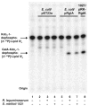
FIGURE 2. A Rhizobiaceae membrane-lipid component is required for RgtA activity in E. coli
Membranes of E. coli harboring pRgtA or the vector control pET23a were assayed under standard conditions at 0.25 mg/ml with or without the addition of 0.25 mg of total lipids extracted from S. meliloti 1021 or R. leguminosarum 3841, as indicated. GalA addition was judged by the shift of the Kdo2-1-dephospho-[4′-32P]lipid IVA band. Membranes of S. meliloti 1021/pRK-RgtA without added lipids were used as the positive control (lane 8).
FIGURE 3
FIGURE 3. Ability of DEAE-cellulose fractionated R. leguminosarum 3841 lipids to support RgtA activity
Total lipids were extracted from R. leguminosarum 3841 and fractionated by DEAE-cellulose column chromatography. Lipids were eluted from the column with the solvent chloroform/methanol/aqueous ammonium acetate (2:3:1, v/v) by increasing the ammonium acetate concentration from 0 to 480 mM in the aqueous component. Lipids (0.25 mg) from each fraction were used in the reconstitution assay. NE, no enzyme; FT, flow-through.
FIGURE 4
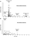
FIGURE 4. Negative ion ESI/MS spectrum of the DEAE-cellulose fraction containing the donor substrate for RgtA
The full spectrum of the R. leguminosarum lipids eluting in the 60 mM fraction from the DEAE-cellulose column is shown in panel A. The full spectrum of the base-stable lipids, eluting in the 60 mM fraction from a second DEAE-cellulose column, is shown in panel B. amu, atomic mass units.
FIGURE 5
FIGURE 5. ESI/MS/MS analysis of the putative GalA donor substrate for RgtA
The MS/MS analysis of the predominant [M – 2H]2− ion at m/z 544.3 in Fig. 4, panel A, is shown together with the proposed fragment ion assignments and the covalent structure of the intact lipid. The proposed stereochemistry of the GalA linkage has not been established by NMR but is proposed based on the sequence similarity of the Rgt proteins to ArnT. amu, atomic mass units.
FIGURE 6
FIGURE 6. Identification of GalA in the dodecaprenyl phosphate-linked HexA isolated from R. leguminosarum
Trimethylsilyl ester derivatives of methyl glycosides, obtained by methanolic HCl hydrolysis of the purified dodecaprenyl phosphate-linked HexA, were separated by gas-liquid chromatography. Peaks were identified by comparison of retention times with standards prepared in parallel. The chromatogram for the lipid donor was compared with those of GalA (see asterisks) and GlcA standards. No GlcA peaks are present in the natural product.
FIGURE 7
FIGURE 7. In vitro reconstitution of RgtA activity with purified dodecaprenyl phosphate-GalA
RgtA was assayed using 2.5 μM of Kdo2-1-dephospho-[4′-32P]lipid IVA as the acceptor substrate under standard conditions for 5 min with 0.25 mg/ml membrane protein. The dodecaprenyl phosphate-GalA was dissolved in the same assay buffer, except that the Triton X-100 concentration was increased to 0.2% to dissolve the purified lipid. Membranes from E. coli NovaBlue (DE3) harboring pRgtA or the vector pET23a were used as indicated. The purified donor substrate was added in increasing concentrations, ranging from 0.1 to 100 μM. The reconstituted E. coli system was run alongside S. meliloti 1021/pRgtA control membranes.
FIGURE 8
FIGURE 8. In vitro reconstitution of RgtA, RgtB, and RgtC activities with purified dodecaprenyl phosphate-GalA
RgtA, RgtB, and RgtC were assayed using mannosyl-Kdo2-1-dephospho-[4′-32P]lipid IVA as the acceptor substrate (no carrier) under standard assay conditions for 30 min with 0.25 mg/ml of each membrane protein. Membranes from E. coli NovaBlue (DE3) harboring pRgtA, pRgtB, pRgtC, or the vector pET23a were used as indicated. The purified donor substrate was added at 200 μM. The three metabolites (products I, II, and III′) seen in the original clone (pMKGE) can be generated by the sequential action of RgtA, RgtB, and RgtC expressed in E. coli, when supplied with the purified GalA donor.
FIGURE 9
FIGURE 9. Proposed pathway and topography for attachment of GalA residues to the lipid A and core domains of R. leguminosarum LPS
The simplest scenario for the biosynthesis of dodecaprenyl-β-D-GalA is as follows. and , UDP-Glc is oxidized by the dehydrogenase Exo5 to UDP-GlcA, which is then converted by the C4-epimerase LpsL to UDP-GalA (22). , in analogy to PmrF (ArnC) (30) in E. coli, Orf3 transfers GalA from UDP-GalA to dodecaprenyl phosphate, generating dodecaprenyl phosphate-β-D-GalA, which is flipped to the periplasmic leaflet by an unknown mechanism. As yet, we have not been able to assay Orf3 in vitro. 4, lipid A and core LPS sugars are synthesized on the cytoplasmic leaflet of the inner membrane (–46, 56, 57) and flipped to the periplasmic leaflet by MsbA. and , on the periplasmic side of the inner membrane the 1- and 4′-phosphatases, LpxE and LpxF (40, 50), dephosphorylate lipid A, creating the substrate for GalA addition. –, GalA is transferred from dodecaprenyl-β-D-GalA to the outer Kdo by RgtA and RgtB, and to mannose by RgtC. The same GalA donor is presumably used in the modification of lipid A by RgtD, but an in vitro assay is not yet available.

Similar articles

Cited by

References

    1. Bugg TD, Brandish PE. FEMS Microbiol Lett. 1994;119:255–262. - PubMed
    1. Lazar K, Walker S. Curr Opin Chem Biol. 2002;6:786–793. - PubMed
    1. Guan Z, Breazeale SD, Raetz CRH. Anal Biochem. 2005;345:336–339. - PubMed
    1. Kelleher DJ, Karaoglu D, Gilmore R. Glycobiology. 2001;11:321–333. - PubMed
    1. Raetz CRH, Whitfield C. Annu Rev Biochem. 2002;71:635–700. - PMC - PubMed

Publication types

LinkOut - more resources