. 2006 Jun;147(6):3007-15.
doi: 10.1210/en.2005-1239. Epub 2006 Feb 23.

Directed differentiation of mouse embryonic stem cells into thyroid follicular cells

Affiliations

Directed differentiation of mouse embryonic stem cells into thyroid follicular cells

Maria C Arufe et al. Endocrinology. 2006 Jun.

Abstract

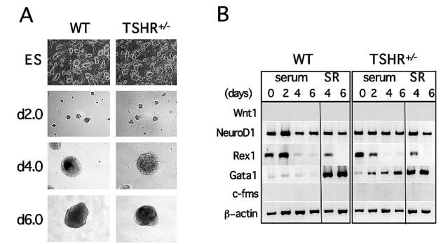
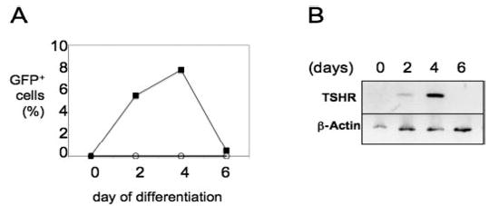
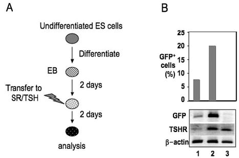
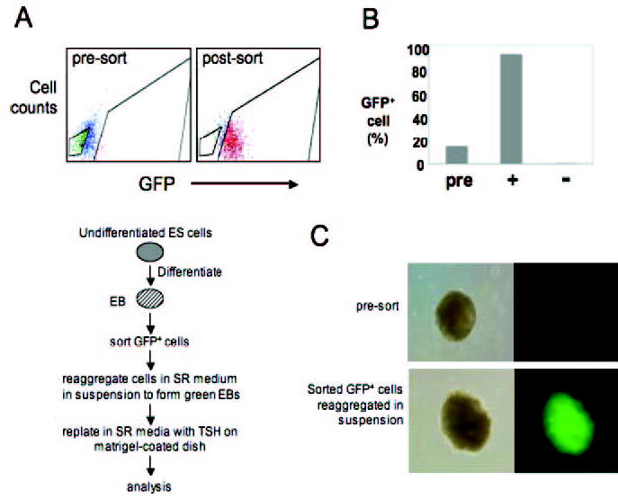
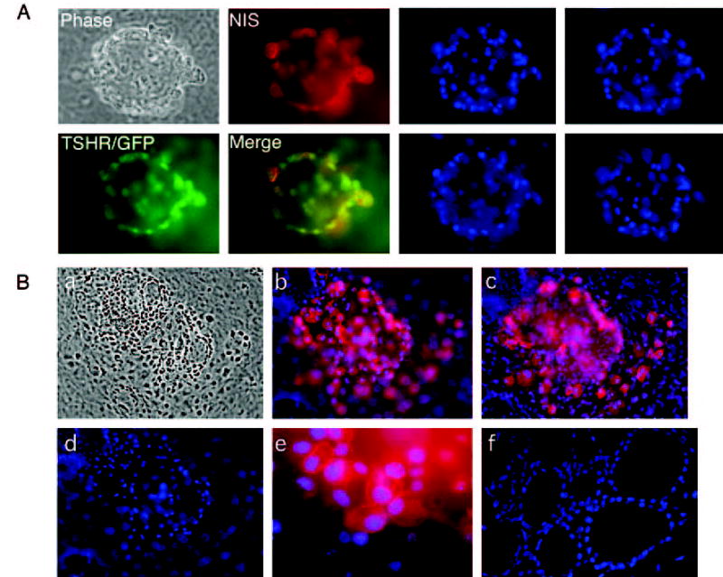
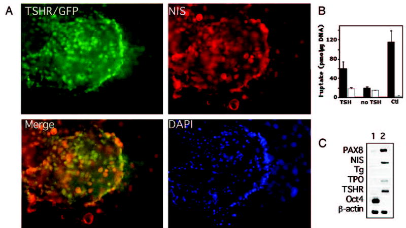
Elucidating the molecular mechanisms leading to the induction and specification of thyroid follicular cells is important for our understanding of thyroid development. To characterize the key events in this process, we previously established an experimental embryonic stem (ES) cell model system, which shows that wild-type mouse CCE ES cells can give rise to thyrocyte-like cells in vitro. We extend our analysis in this report by using a genetically manipulated ES cell line in which green fluorescent protein (GFP) cDNA is targeted to the TSH receptor (TSHR) gene, linking GFP expression to the transcription of the endogenous TSHR gene. The appearance of GFP-positive cells was dependent on the formation of embryoid bodies from undifferentiated ES cells and was greatly enhanced by TSH treatment during the first 2-4 d of differentiation. With the support of Matrigel, highly enriched ES cell-derived GFP-positive cells formed thyroid follicle-like clusters in a serum-free medium supplemented with TSH. Importantly, these clusters display the characteristics of thyroid follicular cells. Immunofluorescent studies confirmed the colocalization of TSHR with the Na+/I- symporter in the clusters and indicated that Na+/I- symporter was expressed exclusively in the plasma membrane. In addition, I- uptake activity was observed in these cells. Our results indicate that ES cells can be induced to differentiate into thyroid follicular cells, providing a powerful tool to study embryonic thyroid development and function.


Figures

Fig. 1
Fig. 1
Characterization of TSHR+/− ES cells. A, Phase contrast micrographs of undifferentiated ES cells and differentiated EBs show that TSHR+/− ES cells and EBs were indistinguishable from their wild-type (WT) counterparts with respect to morphology and size. Numbers on the left indicate days of differentiation. Images were taken with an ECLIPSE TE200-S camera (Nikon Corp., Melville, NY) at × 10 magnification. B, RT-PCR expression analysis of EBs differentiated in serum-containing (serum) medium or in serum-containing medium followed by SR medium for various markers characteristics of primary germ layers, including Wnt1, NeuroD, Gata1, and c-fms. Rex1 is a stem cell marker. Numbers on top of the figure indicate days of differentiation.
Fig. 2
Fig. 2
Kinetics of GFP and TSHR expression in early TSHR+/− EBs. A, Percentage of GFP-positive cells during the first 6 d of culture in EBDM. ○, Wild-type EBs; ▪, TSHR+/− EBs. B, Kinetics of TSHR gene expression patterns during the first 6 d of EB development. Total RNA was isolated from differentiated EBs cultured as described in Materials and Methods. cDNA was analyzed from total RNA using oligo(deoxythymidine) primers. RT-PCR was performed to detect the expression of TSHR. β-Actin served as an internal control. Numbers on top of the figure indicate days of differentiation.
Fig. 3
Fig. 3
Effects of TSH on GFP and TSHR expression. A, General outline of the differentiation protocol. B, Upper panel, Flow cytometric analysis of GFP expression in TSHR+/− EBs differentiated in SR medium (lane 1) or SR medium supplemented with hTSH (lane 2). Note that GFP expression was substantially induced in TSH-supplemented cells cultured in SR medium. Wild-type EBs were used as negative controls (lane 3). Lower panel, RT-PCR expression analysis demonstrating the effect of TSH on TSHR and GFP gene expression in d 4 EBs.
Fig. 4
Fig. 4
Selective enrichment of GFP-expressing cells. A, FACS analysis of d 4 EBs and culture protocol for differentiation of sorted GFP+ cells. B, FACS analysis of cells from the presorted (pre), GFP-positive (+), and GFP-negative (−) populations. C, Immunofluorescent analysis demonstrates the appearance of green EBs in reaggregated, sorted GFP+ cells in suspension. Note that presorted EBs do not show detectable levels of fluorescence. Left panels, Phase contrast images; right panels, fluorescent microscopic images.
Fig. 5
Fig. 5
GFP+NIS+ cells are organized into one or more follicle-like cell clusters. Left panel, A follicle-like cluster derived from TSHR+/− ES cells after 21 d of differentiation visualized microscopically (phase) or after staining with an antibody to NIS (red). TSHR expression is indicated by the green GFP signal. An overlaid image shows the colocalization of NIS with TSHR (yellow). Right panel, The same field of cells after nuclear staining with DAPI (blue). Images were taken from different optical sections. B, Immunofluorescent images of several follicle-like clusters derived from TSHR+/− ES cells after 21 d of differentiation. a, Phase contrast exposure. b and c, Immunofluorescent staining, demonstrating the expression of NIS protein. Note that a rim of cells several layers thick adhered closely to the NIS-positive cells. d, Nuclear DAPI staining. e, High magnification of anti-NIS immunofluorescence, demonstrating the expression of NIS in the plasma membrane. Immunofluorescence was not detected when the experiment was performed with the second antibody alone (data not shown). f, Nuclear DAPI staining of native mouse thyroid tissue.
Fig. 6
Fig. 6
Thyroid potential of GFP+NIS+ cells. A, Several clusters of GFP+NIS+ cells derived from TSHR+/− ES cells after staining with an antibody to NIS (red). In merging, some areas showed overlay (yellow). Blue indicates nuclear DAPI staining. B, I uptake activity at 20 μM external sodium iodide in TSHR+/− ES cells after 21 d of differentiation. ▪, Absence of 40 μM NaClO4; □, presence of 40 μM NaClO4. Note that cells treated with TSH showed I uptake activity, whereas cells maintained in the absence of TSH did not. I uptake in NIS/MDCK cells (Ctl) was measured as a positive control for functional NIS activity. The values represent the mean ± SE of two independent experiments performed in triplicate. C, Gene expression analysis by RT-PCR shows differentiation of thyroid follicular cells from ES cells. RNA was isolated from undifferentiated ES cells (lane 1) and thyroid follicular cells grown for 21 d (lane 2) and was analyzed for the expression of thyroid marker genes PAX8, NIS, Tg, TPO, and TSHR. Oct4 is an undifferentiated ES cell marker. β-Actin served as an internal control standard.

Similar articles

Cited by

References

    1. Ambesi-Impiombato FS, Coon HG. Thyroid cells in culture. Int Rev Cytol. 1979;(Suppl):163–172. - PubMed
    1. Fayet G, Hovsepian S. Isolation of a normal human thyroid cell line: hormonal requirement for thyroglobulin regulation. Thyroid. 2002;12:539–546. - PubMed
    1. Damante G, Tell G, Di Lauro R. A unique combination of transcription factors controls differentiation of thyroid cells. Prog Nucleic Acids Res Mol Biol. 2001;66:307–356. - PubMed
    1. Tasevski V, Benn D, Peters G, Luttrell B, Simpson A. The Fischer rat thyroid cell line FRTL-5 exhibits a nondiploid karyotype. Thyroid. 1998;8:623–626. - PubMed
    1. Ealey PA, Emmerson JM, Bidey SP, Marshall NJ. Thyrotrophin stimulation of mitogenesis of the rat thyroid cell strain FRTL-5: a metaphase index assay for the detection of thyroid growth stimulators. J Endocrinol. 1985;106:203–210. - PubMed

Publication types