

A common phosphotyrosine signature for the Bcr-Abl kinase
- PMID: 16497976
- PMCID: PMC1895816
- DOI: 10.1182/blood-2005-08-3399
A common phosphotyrosine signature for the Bcr-Abl kinase
Abstract
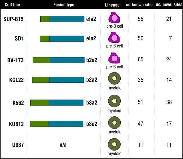
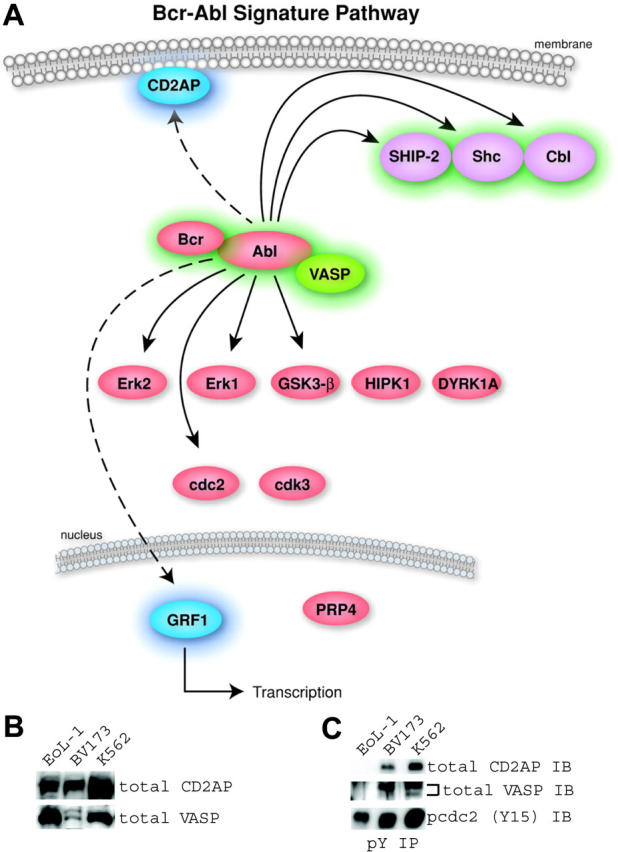
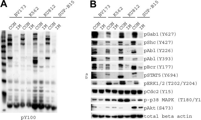
The Bcr-Abl fusion kinase drives oncogenesis in chronic myeloid leukemia (CML). CML patients are currently treated with the Abl tyrosine kinase inhibitor imatinib, which is effective in early stages of the disease. However, resistance to imatinib arises in later disease stages primarily because of a Bcr-Abl mutation. To gain deeper insight into Bcr-Abl signaling pathways, we generated phosphotyrosine profiles for 6 cell lines that represent 3 Bcr-Abl fusion types by using immunoaffinity purification of tyrosine phosphopeptides followed by tandem mass spectrometry. We identified 188 nonredundant tyrosine-phosphorylated sites, 77 of which are novel. By comparing the profiles, we found a number of phosphotyrosine sites common to the 6 cell lines regardless of cellular background and fusion type, several of which are decreased by imatinib treatment. Comparison of this Bcr-Abl signature with the profile of cells expressing an alternative imatinib-sensitive fusion kinase, FIP1L1-PDGFRalpha, revealed that these kinases signal through different pathways. This phosphoproteomic study of the Bcr-Abl fusion kinase highlights novel disease markers and potential drug-responsive biomarkers and adds novel insight into the oncogenic signals driven by the Bcr-Abl kinase.
Figures
References
-
- Sawyers CL. Chronic myeloid leukemia. N Engl J Med. 1999;340: 1330-1340. - PubMed
-
- Deininger MW, Goldman JM, Melo JV. The molecular biology of chronic myeloid leukemia. Blood. 2000;96: 3343-3356. - PubMed
-
- Nowell P, Hungerford DA. A minute chromosome in human chronic granulocytic leukemia. Science. 1960;132: 1497.
-
- Deininger MW, Druker BJ. Specific targeted therapy of chronic myelogenous leukemia with imatinib. Pharmacol Rev. 2003;55: 401-423. - PubMed
-
- O'Dwyer ME, La Rosee P, Nimmanapalli R, Bhalla KN, Druker BJ. Recent advances in Philadelphia chromosome-positive malignancies: the potential role of arsenic trioxide. Semin Hematol. 2002;39: 18-21. - PubMed
Publication types
MeSH terms
Substances
Grants and funding
LinkOut - more resources
Full Text Sources
Other Literature Sources
Medical
Molecular Biology Databases
Miscellaneous
