. 2006 Mar 17;98(5):e39-47.
doi: 10.1161/01.RES.0000215436.92414.1d. Epub 2006 Feb 23.

Extracellular nucleotides and adenosine independently activate AMP-activated protein kinase in endothelial cells: involvement of P2 receptors and adenosine transporters

Affiliations

Extracellular nucleotides and adenosine independently activate AMP-activated protein kinase in endothelial cells: involvement of P2 receptors and adenosine transporters

Cleide Gonçalves da Silva et al. Circ Res. .

Abstract

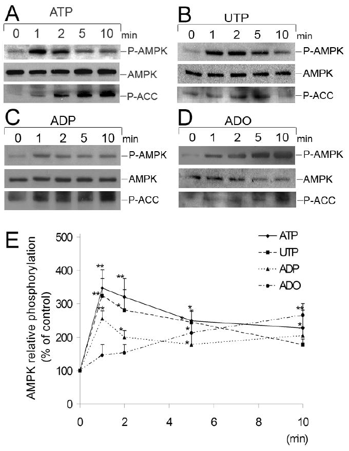
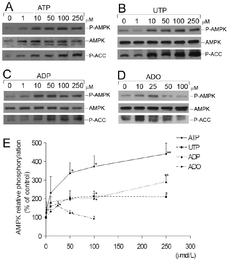
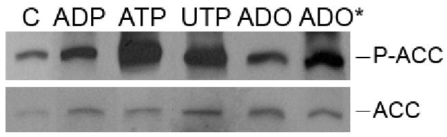
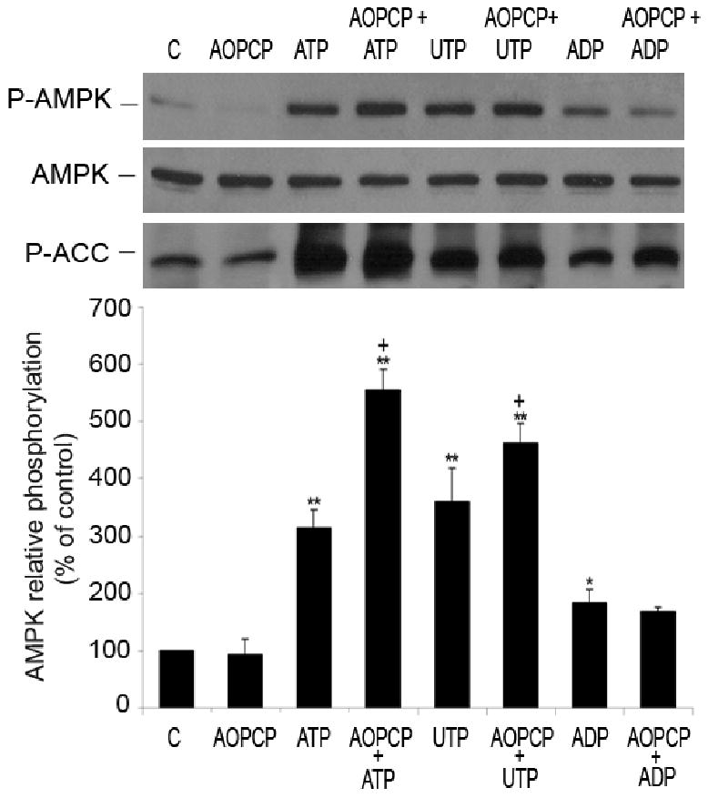
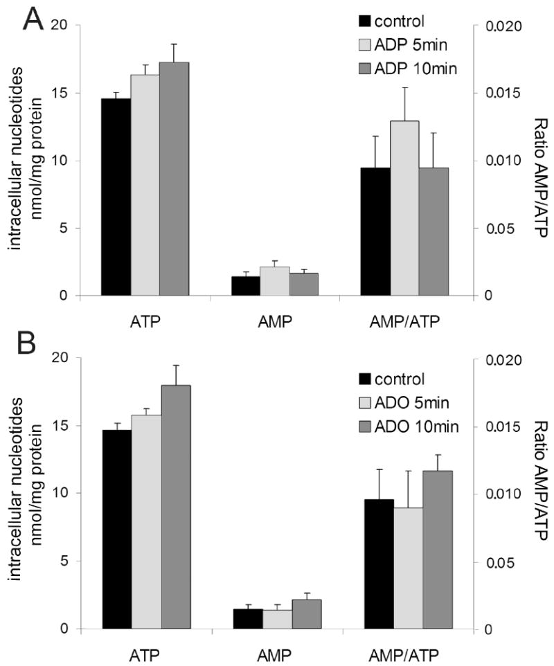
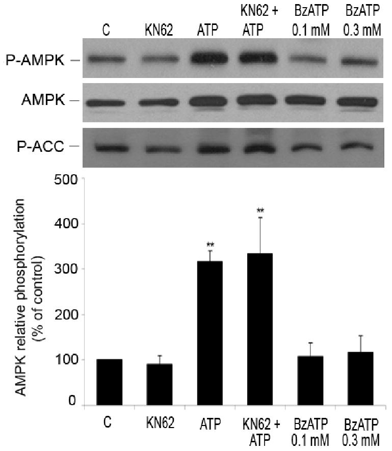
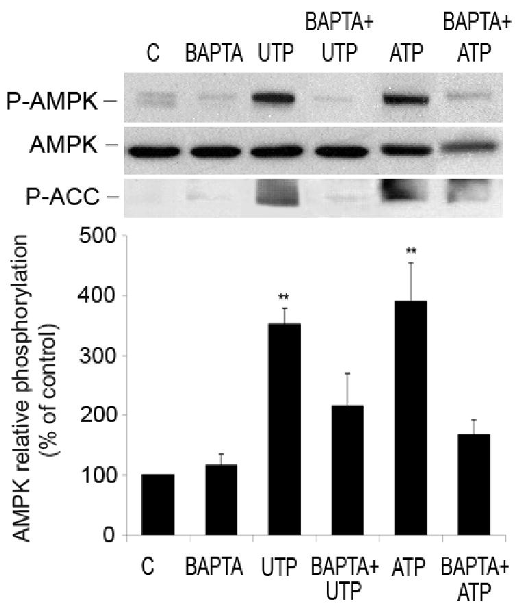
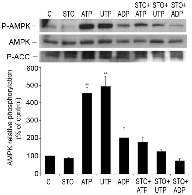
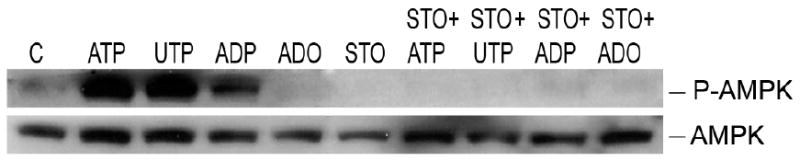
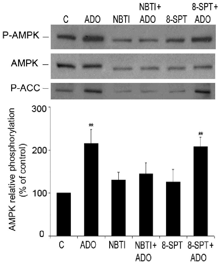
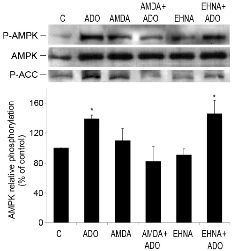
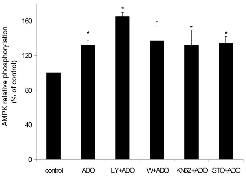
AMP-activated protein kinase (AMPK) plays a key role in the regulation of energy homeostasis and is activated in response to cellular stress, including hypoxia/ischemia and hyperglycemia. The stress events are accompanied by rapid release of extracellular nucleotides from damaged tissues or activated endothelial cells (EC) and platelets. We demonstrate that extracellular nucleotides (ATP, ADP, and UTP, but not UDP) and adenosine independently induce phosphorylation and activation of AMPK in human umbilical vein EC (HUVEC) by the mechanism that is not linked to changes in AMP:ATP ratio. HUVEC express NTPDases, as well as 5'-nucleotidase; hence, nucleotides can be metabolized to adenosine. However, inhibition of 5'-nucleotidase had no effect on ATP/ADP/UTP-induced phospho- rylation of AMPK, indicating that AMPK activation occurred as a direct response to nucleotides. Nucleotide-evoked phosphorylation of AMPK in HUVEC was mediated by P2Y1, P2Y2, and/or P2Y4 receptors, whereas P2Y6, P2Y11, and P2X receptors were not involved. The nucleotide-induced phosphorylation of AMPK was affected by changes in the concentration of intracellular Ca2+ and by Ca2+/calmodulin-dependent kinase kinase (CaMKK), although most likely it was not dependent on LKB1 kinase. Adenosine-induced phosphorylation of AMPK was not mediated by P1 receptors but required adenosine uptake by equilibrative nucleoside transporters followed by its (intracellular) metabolism to AMP. Moreover, adenosine effect was Ca2+ and CaMKK independent, although probably associated with upstream LKB1. We hypothesize that P2 receptors and adenosine transporters could be novel targets for the pharmacological regulation of AMPK activity and its downstream effects on EC function.


Figures

Figure 1
Figure 1
Time-course studies of phosphorylation of AMPK induced by nucleotides and adenosine in HUVEC. Representative Western blots of HUVEC stimulated by (A) ATP 100 μmol/L, (B) UTP 100 μmol/L, (C) ADP 100 μmol/L or (D) adenosine (ADO) (25 μmol/L) from 0 to 10 minutes. Cell lysates were immunoblotted with anti-phospho-AMPK (Thr-172) (P-AMPK), anti-AMPK-α and anti-P-ACC antibodies. (E) Results are expressed as percentage of the control (0 minutes) AMPK relative phosphorylation. Values are presented as means ± S.E.M of 3-5 independent experiments. *P<0.05, ** P< 0.01 compared to the control.
Figure 2
Figure 2
Concentration-dependence of nucleotide- and adenosine-induced phosphorylation of AMPK in HUVEC. Representative Western blots of HUVEC stimulated for 5 minutes with the indicated concentrations of (A) ATP, (B) UTP, (C) ADP or (D) adenosine (ADO), ranging from 0 to 250 μmol/L. Cell lysates were immunoblotted with anti-P-AMPK (Thr-172), anti-AMPK-α and anti-P-ACC antibodies. (E) Results are expressed as percentage of the control (0 μmol/L) AMPK relative phosphorylation. Values are means ± S.E.M of 3-4 independent experiments. *P<0.05, **P< 0.01 compared to control.
Figure 3
Figure 3
Extracellular nucleotides and adenosine induce phosphorylation of acetyl-CoA carboxylase (ACC). Western blot analysis of HUVEC stimulated by ADP (100 μmol/L), ATP (100 μmol/L), UTP (100 μmol/L) or adenosine (ADO) (25 μmol/L) for 5 minutes or ADO* for 30 minutes. Cell lysates were immunoblotted with anti-phospho-ACC (Ser-79) (P-ACC) or Streptavidin-HRP antibody (total ACC). Results are representative of 4-5 independent experiments.
Figure 4
Figure 4
Nucleotide-induced phosphorylation of AMPK is not adenosine-dependent. Representative Western blot of HUVEC pre-treated for 15 minutes with inhibitor of 5′nucleotidase, AOPCP (100 μmol/L) and incubated with or without nucleotides (100 μmol/L, 5 minutes). Cell lysates were immunoblotted with anti-P-AMPK (Thr-172), anti-AMPK-α and anti-P-ACC antibodies. After quantification, values were statistically analyzed and are expressed as means ± S.E.M of 2-4 independent experiments. *P<0.05, **P< 0.01 compared to control, +P< 0.05 compared to non pre-incubated cells, treated with respective nucleotide.
Figure 5
Figure 5
Changes in AMP:ATP ratio do not affect AMPK activation by extracellular nucleotides and adenosine. HUVEC were stimulated with (A) 100 μmol/L ADP or (B) 25 μmol/L adenosine for 5 and 10 minutes and concentrations of intracellular AMP and ATP were measured by HPLC. Data are expressed as means ± S.E.M of 3 independent experiments. P>0.05 compared to control.
Figure 6
Figure 6
Role of P2X and P2Y11 receptors in AMPK phosphorylation. HUVEC were stimulated with ATP (1 mmol/L, 5 minutes) with and without pre-incubation with KN62 (1 μmol/L, 20 minutes), the antagonist of P2X7 receptor. Cells were also treated with BzATP (100 and 300 μmol/L, 5 minutes), an agonist for P2X and P2Y11 receptors. Cell lysates were analyzed by Western blotting using antibodies to P-AMPK (Thr-172), AMPK-α and P-ACC. Results obtained from 3 independent experiments are presented as means ± S.E.M. **P< 0.01 compared to control.
Figure 7
Figure 7
Phosphorylation of AMPK induced by nucleotides depends on Ca2+ originating from intracellular stores. Representative Western blots showing the effect of BAPTA-AM on nucleotide-induced AMPK and ACC phosphorylation. HUVEC were pre-treated with BAPTA-AM (20 minutes, 10 μmol/L) and stimulated with ATP and UTP (1 minute, 100 μmol/L). Cell lysates were immunoblotted with antibodies to P-AMPK, AMPK-α and P-ACC. After quantification, values were statistically analyzed and are expressed as means ± S.E.M of 3-6 independent experiments. *P < 0.05 and **P < 0.01 compared to untreated cells.
Figure 8
Figure 8
Nucleotide-induced phosphorylation of AMPK is mediated by CAMKK in HUVEC. Representative Western blot of HUVEC pre-treated for 30 minutes with STO-609 (1μg/μL), inhibitor of CAMKK, and incubated with or without nucleotides (100 μmol/L, 1 minute). Cell lysates were immunoblotted with anti-P-AMPK (Thr-172), anti-AMPK-α and anti-P-ACC antibodies. After quantification, values were statistically analyzed and are expressed as means ± S.E.M of 3 independent experiments. *P<0.05, **P< 0.01 compared to control.
Figure 9
Figure 9
Role of LKB1 in nucleotide and adenosine-mediated activation of AMPK. HeLa cells were incubated with extracellular nucleotides, ATP, UTP and ADP (100 ìmol/L; 2 minutes) or adenosine (25 ìmol/L; 5 minutes). Representative Western blots show cell lysates immunoblotted with antibodies to P-AMPK (Thr-172) and AMPK-α. Representative Western blots of 3 independent experiments.
Figure 10
Figure 10
Adenosine transporters and not P1 receptors are involved in adenosine-induced phosphorylation of AMPK. Representative Western blots of HUVEC pre-treated for 20 minutes with either an antagonist of P1 receptors, 8-SPT (30 μmol/L), or inhibitor of adenosine transporter NBTI (10 μmol/L), and incubated with adenosine (ADO) (25 μmol/L, 5 minutes). Cell lysates were immunoblotted with antibodies to P-AMPK (Thr-172), AMPK-α and P-ACC. After quantification, values were statistically analyzed and are expressed as means ± S.E.M of 5-7 independent experiments. **P< 0.01 compared to the control.
Figure 11
Figure 11
Intracellular metabolism of adenosine to AMP is essential for phosphorylation of AMPK. Representative Western blots of HUVEC pre-treated for 30 minutes with either an inhibitor of adenosine kinase, AMDA (10 μmol/L) or inhibitor of adenosine deaminase, EHNA (10 μmol/L), and stimulated with adenosine (ADO) (25 μmol/L, 5 minutes). Cell lysates were immunoblotted with anti-P-AMPK (Thr-172), anti-AMPK-α and anti-P-ACC antibodies. Values are means ± S.E.M of 3-4 independent experiments. *P<0.05, compared to control.
Figure 12
Figure 12
Adenosine-induced AMPK phosphorylation is independent on CaMK II, CaMKK and PI3K. HUVEC were pre-treated for 20 minutes with inhibitors of PI3K (LY294002, 10 μmol/L or wortmannin (W); 100 nmol/L), CaMK II (KN62, 10 μmol/L) and CaMKK (STO-609, 1 μg/μL) and stimulated with adenosine (ADO) (25 μmol/L, 5 minutes). Cell lysates were immunoblotted with anti-P-AMPK (Thr-172) and anti-AMPK-α antibodies and the bands were densitometrically quantified. Results are expressed as means ± S.E.M of 3-4 independent experiments. *P<0.05, compared to control.

Similar articles

Cited by

References

    1. Hardie DG, Salt IP, Hawley SA, Davies SP. AMP-activated protein kinase: an ultrasensitive system for monitoring cellular energy charge. Biochem J. 1999;338(Pt 3):717–22. - PMC - PubMed
    1. Hardie DG, Salt IP, Davies SP. Analysis of the role of the AMP-activated protein kinase in the response to cellular stress. Methods Mol Biol. 2000;99:63–74. - PubMed
    1. Daniel T, Carling D. Expression and regulation of the AMP-activated protein kinase-SNF1 (sucrose non-fermenting 1) kinase complexes in yeast and mammalian cells: studies using chimaeric catalytic subunits. Biochem J. 2002;365:629–38. - PMC - PubMed
    1. Hawley SA, Davison M, Woods A, Davies SP, Beri RK, Carling D, Hardie DG. Characterization of the AMP-activated protein kinase kinase from rat liver and identification of threonine 172 as the major site at which it phosphorylates AMP-activated protein kinase. J Biol Chem. 1996;271:27879–87. - PubMed
    1. Stein SC, Woods A, Jones NA, Davison MD, Carling D. The regulation of AMP-activated protein kinase by phosphorylation. Biochem J. 2000;345(Pt 3):437–43. - PMC - PubMed

Publication types

MeSH terms