. 2006 Mar;85(3):267-71.
doi: 10.1177/154405910608500312.

Novel MSX1 frameshift causes autosomal-dominant oligodontia

Affiliations

Novel MSX1 frameshift causes autosomal-dominant oligodontia

J-W Kim et al. J Dent Res. 2006 Mar.

Abstract

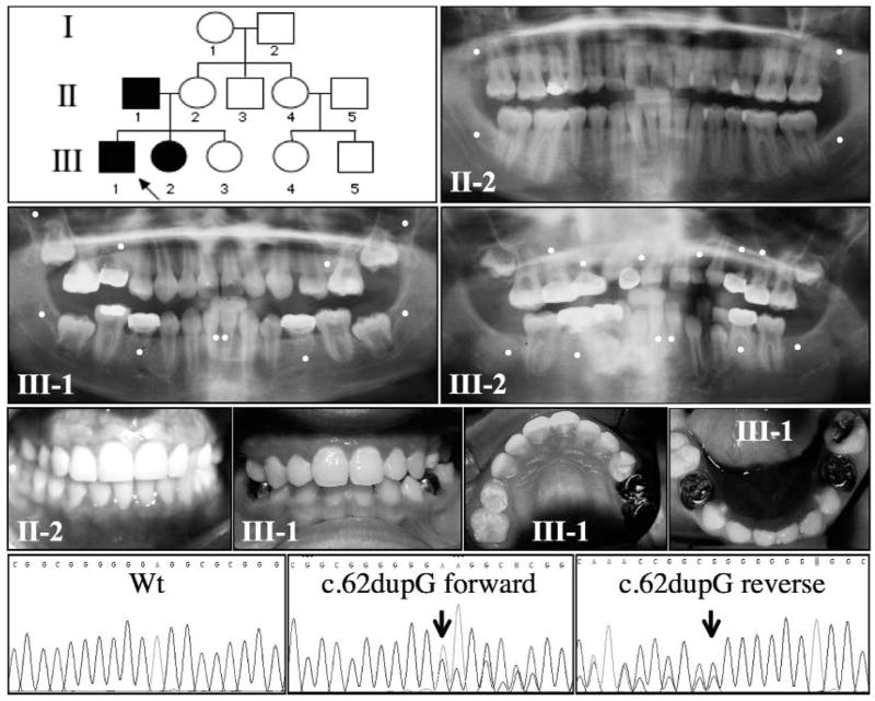
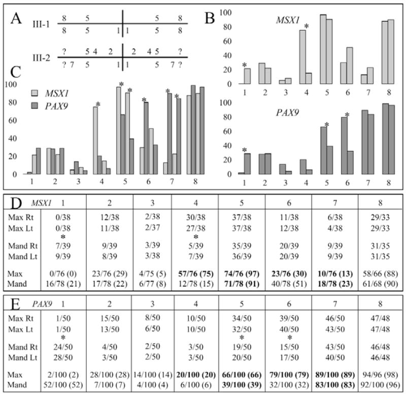
Can kindreds with tooth agenesis caused by MSX1 or PAX9 mutations be distinguished by their phenotypes? We have identified an MSX1second bicuspids and mandibular central incisors. The dominant phenotype is apparently due to haploinsufficiency. We analyzed patterns of partial tooth agenesis in seven kindreds with defined MSX1 mutations and ten kindreds with defined PAX9 mutations. The probability of missing a particular type of tooth is always bilaterally symmetrical, but differences exist between the maxilla and mandible. MSX1-associated oligodontia typically includes missing maxillary and mandibular second bicuspids and maxillary first bicuspids. The most distinguishing feature of MSX1-associated oligodontia is the frequent (75%) absence of maxillary first bicuspids, while the most distinguishing feature of PAX9-associated oligodontia is the frequent (> 80%) absence of the maxillary and mandibular second molars.


Figures

Figure 1
Figure 1
Oral photographs of the unaffected mother (II-2) and the proband (III-1; age 11) show that the MSX1 mutation (g.62dupG) has no apparent effect on tooth size (top). Panorex radiographs demonstrate that the unaffected mother (II-2) has all of her permanent teeth except the third molars, while the proband (III-1) is missing 6 teeth, and his affected sister (III-2; age 9) is missing 12 teeth, not counting the third molars. A white dot indicates the expected location of a congenitally absent tooth. The pedigree indicates that the oligodontia trait in this family was transmitted from the father to the offspring in an autosomal-dominant pattern of inheritance. DNA sequencing chromatograms (bottom) identified the specific MSX1 frameshift mutation (arrows) in both the forward and reverse directions. This mutation was not observed in the wild-type (Wt) MSX1 gene in the unaffected members of the kindred.
Figure 2
Figure 2
Quadrant diagram (Palmer notation system) indicating the teeth missing in the two affected members (III-1 and III-2) of the MSX1 kindred (A). The number of missing teeth in people with defined MSX1 mutations was compiled for each position in the human dentition (32 teeth) based on the data from this report, as well as from the six previous reports of MSX1 mutations. The MSX1 mutations are summarized in the chart (D) as fractions, with the numerators being the number of missing teeth and the denominators being the number of individuals. There were no statistically significant differences between the numbers of teeth missing on the left and right, so the data for equivalent teeth on the left and right were combined (bottom of D). We plotted the percentage of teeth missing for each position in the maxillary and mandibular arches (B top), and indicated statistically significant differences (p < 0.001) between the maxillary and mandibular arches with asterisks. The number of missing teeth at each position in people with defined PAX9 mutations was compiled from the 8 previous reports of 10 PAX9 mutations (E). As with MSX1, there were no statistically significant differences between the numbers of teeth missing on the left and right, so the data for equivalent teeth on the left and right were combined (bottom of E). In subsequent analyses, we averaged the values for the mandibular central and lateral incisors, since which tooth is missing is often ambiguous. We plotted the percentage of teeth missing for each position in the maxillary and mandibular arches (B bottom), with statistically significant differences (p < 0.001) between the maxillary and mandibular arches marked with asterisks. We plotted the percentage of missing teeth at each maxillary and mandibular position for people with defined MSX1 and PAX9 mutations (C). For each tooth position in the bar graphs (B,C), the bars for the maxillary teeth are on the right, and bars for the mandibular teeth are on the left. Statistically significant differences (p < 0.001) between MSX1 and PAX9 are marked with asterisks in C, and by bold type in D and E. Note that the absence of maxillary first bicuspids (#4) is the most distinguishing feature of an MSX1 mutation, while the absence of second molars (#7) is the most distinguishing feature of PAX9 mutations. Key: 1, central incisor; 2, lateral incisor; 3, cuspid; 4, first bicuspid; 5, second bicuspid; 6, first molar; 7, second molar 8, third molar; ?, not able to be determined; x axis = tooth position; y axis = percent missing.

References

    1. Bendall AJ, Rincon-Limas DE, Botas J, Abate-Shen C. Protein complex formation between Msx1 and Lhx2 homeoproteins is incompatible with DNA binding activity. Differentiation. 1998;63:151–157. - PubMed
    1. Bendall AJ, Ding J, Hu G, Shen MM, Abate-Shen C. Msx1 antagonizes the myogenic activity of Pax3 in migrating limb muscle precursors. Development. 1999;126:4965–4976. - PubMed
    1. Chen Y, Bei M, Woo I, Satokata I, Maas R. Msx1 controls inductive signaling in mammalian tooth morphogenesis. Development. 1996;122:3035–3044. - PubMed
    1. Das P, Stockton DW, Bauer C, Shaffer LG, D'Souza RN, Wright T, et al. Haploinsufficiency of PAX9 is associated with autosomal dominant hypodontia. Hum Genet. 2002;110:371–376. - PubMed
    1. Das P, Hai M, Elcock C, Leal SM, Brown DT, Brook AH, et al. Novel missense mutations and a 288-bp exonic insertion in PAX9 in families with autosomal dominant hypodontia. Am J Med Genet A. 2003;118:35–42. - PMC - PubMed

Publication types