. 2006 Mar 8;25(5):977-86.
doi: 10.1038/sj.emboj.7601008. Epub 2006 Feb 23.

ICER induced by hyperglycemia represses the expression of genes essential for insulin exocytosis

Affiliations

ICER induced by hyperglycemia represses the expression of genes essential for insulin exocytosis

Amar Abderrahmani et al. EMBO J. .

Abstract

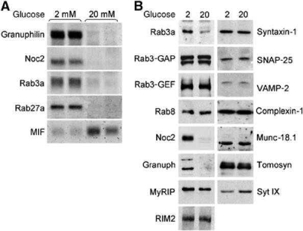
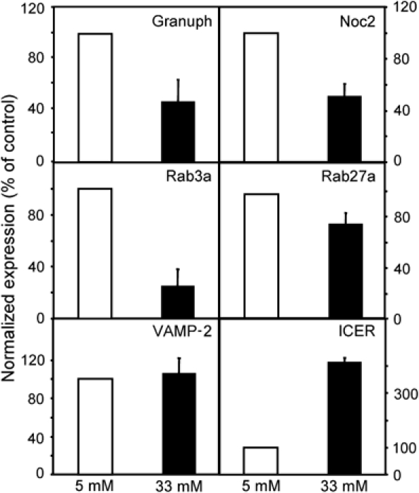
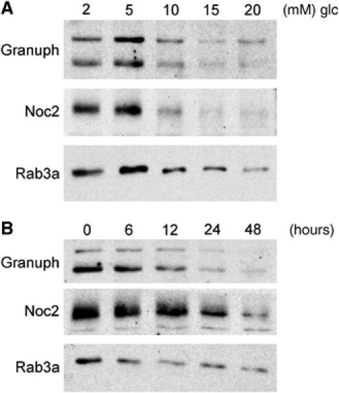
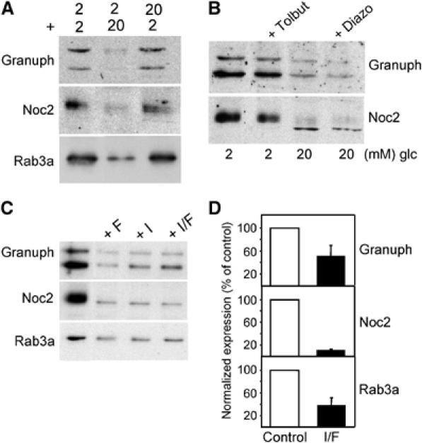
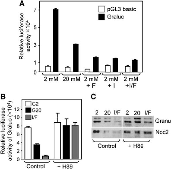
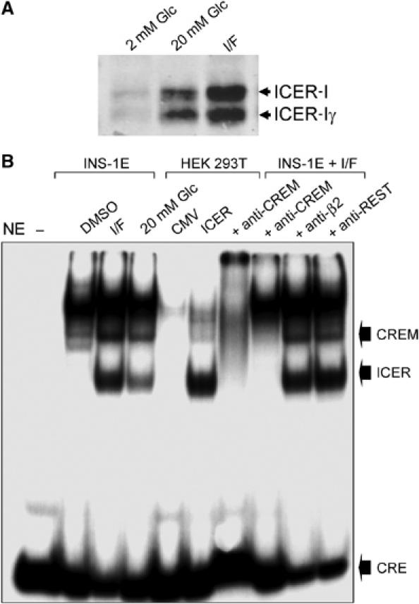
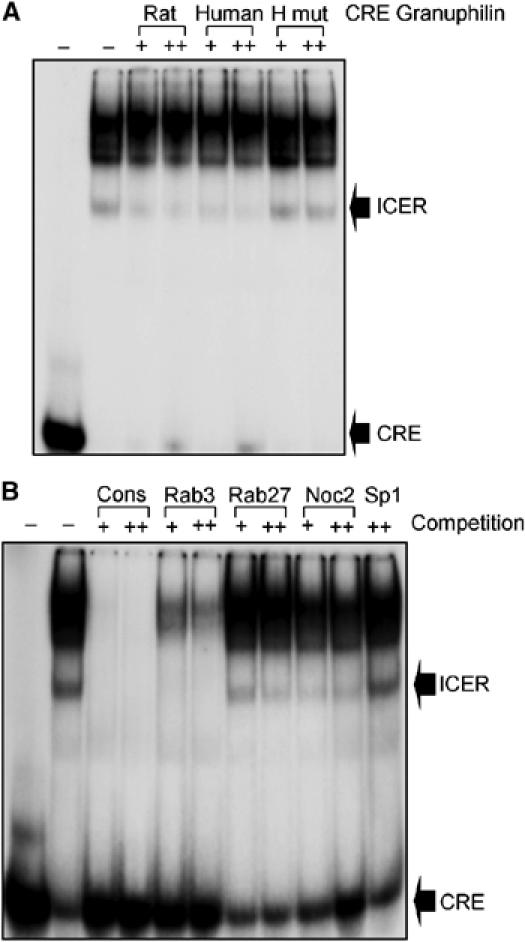
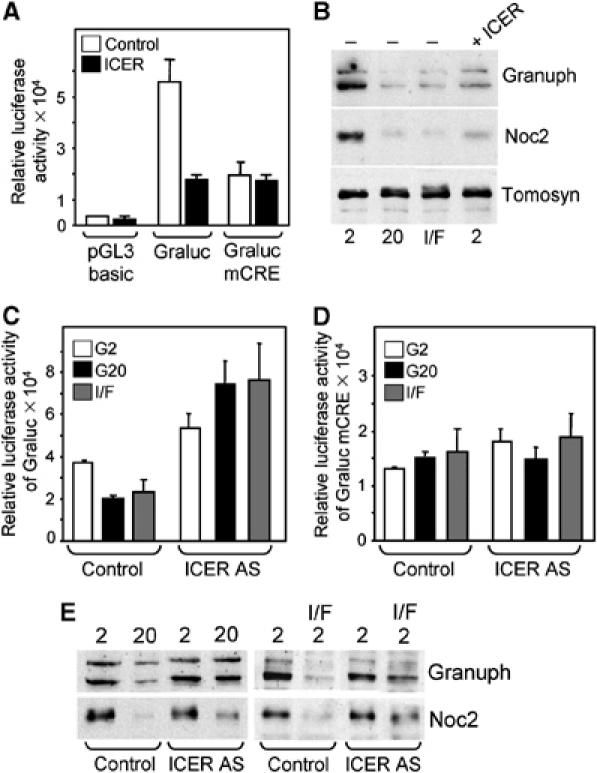
The GTPases Rab3a and Rab27a and their effectors Granuphilin/Slp4 and Noc2 are essential regulators of neuroendocrine secretion. Chronic exposure of pancreatic beta-cells to supraphysiological glucose levels decreased selectively the expression of these proteins. This glucotoxic effect was mimicked by cAMP-raising agents and blocked by PKA inhibitors. We demonstrate that the transcriptional repressor ICER, which is induced in a PKA-dependent manner by chronic hyperglycemia and cAMP-raising agents, is responsible for the decline of the four genes. ICER overexpression diminished the level of Granuphilin, Noc2, Rab3a and Rab27a by binding to cAMP responsive elements located in the promoters of these genes and inhibited exocytosis of beta-cells in response to secretagogues. Moreover, the loss in the expression of the genes of the secretory machinery caused by glucose and cAMP-raising agents was prevented by an antisense construct that reduces ICER levels. We propose that induction of inappropriate ICER levels lead to defects in the secretory process of pancreatic beta-cells possibly contributing, in conjunction with other known deleterious effects of hyperglycemia, to defective insulin release in type 2 diabetes.


Figures

Figure 1
Figure 1
Effect of prolonged exposure to elevated glucose concentrations on mRNA and protein levels of Granuphilin, Noc2, Rab3a and Rab27a. (A) INS-1E cells were cultured in the presence of 2 or 20 mM glucose. After 2 days, total RNA was extracted and the expression of the indicated genes analyzed by Northern blotting. (B) INS-1E cells were cultured for 2 days at 2 or 20 mM glucose. Expression of the indicated proteins involved in the regulation of insulin exocytosis was determined by Western blotting. The figure shows representative results obtained in at least three independent experiments.
Figure 2
Figure 2
Effect of high glucose on the expression of key regulators of insulin exocytosis in rat pancreatic islets. Freshly isolated rat pancreatic islets were cultured for 96 h at 5 (open bars) or 33 mM glucose (filled bars). Expression of the indicated genes was assessed by real-time PCR analysis. The results were normalized with the values obtained for tubulin in the same sample. Normalization using Rab3GAP values gave similar results. The data obtained in control islets were set to 100%. The results are the mean±s.e.m. of four to five independent experiments performed in triplicates. The mRNA levels of Granuphilin, Noc2, Rab3a Rab27a and ICER at 33mM glucose are significantly different (P<0.001, n=4–5, Student's t-test) than in control cells. The expression of VAMP-2 at 5 and 33 mM glucose was not significantly altered.
Figure 3
Figure 3
Dose and time dependency of the effect of glucose on Granuphilin, Noc2 and Rab3a expression. (A) INS-1E cells were cultured for 2 days in the presence of the indicated glucose (glc) concentrations. The level of Granuphilin, Noc2 and Rab3a was assessed by Western blotting. (B) INS-1E cells were incubated for the indicated periods in the presence of 20 mM glucose. The amount of Granuphilin, Noc2 and Rab3a remaining in the cells was estimated by Western blotting.
Figure 4
Figure 4
The effect of glucose is reversible and can be mimicked by cAMP-raising agents but not with pharmacological agents that affect insulin secretion. (A) The cells were incubated either at 2 mM (2) or 20 mM (20) glucose for 36 h. The medium was then replaced and the cells incubated for a second period of 36 h at 2 or 20 mM glucose. At the end of the experiment, the cells were homogenized and the extracts analyzed by Western blotting with the indicated antibodies. (B) The cells were cultured for 2 days at 2 mM glucose in the presence or absence of 100 μM Tolbutamide (Tolbut) or at 20 mM glucose with or without 200 μM Diazoxide (Diazo). The expression of Granuphilin and Noc2 was analyzed by Western blotting. (C) INS-1E cells were cultured for two days at 2 mM glucose with or without Forskolin (10 μM) (F) and IBMX (100 μM) (I). The level of Granuphilin, Noc2 and Rab3a was assessed by Western blotting. (D) Freshly isolated rat pancreatic islets were incubated with (filled bars) or without (open bars) Forskolin (10 μM) (F) and IBMX (100 μM) (I) for 72 h. Expression of Granuphilin, Noc2 and Rab3a was assessed by real-time PCR analysis. The values obtained in control islets were normalized to 100%. The results are the mean±s.e.m. of three independent experiments performed in triplicates.
Figure 5
Figure 5
The effect of glucose on the activity of the Granuphilin promoter is mimicked by cAMP-raising agents and is prevented by the PKA inhibitor H89. (A) INS-1E cells were transiently transfected either with a plasmid encoding a luciferase reporter gene controlled by a minimal promoter (pGL3 basic, open bars) or driven by the human Granuphilin promoter (Graluc, filled bars). After transfection the cells were cultured for 2 days either at 2 mM glucose with or without Forskolin/IBMX or at 20 mM glucose. (B) INS-1E cells transiently transfected with a plasmid encoding a luciferase reporter gene controlled by the human Granuphilin promoter were cultured for 2 days either at 2 mM glucose (G2) with or without Forskolin/IBMX (I/F) or at 20 mM glucose (G20). Half of the cells were treated with the PKA inhibitor H89 (10 μM) while control cells were incubated with vehicle. All results from transfection experiments are the mean±s.e.m. of three independent experiments performed in triplicates. (C) INS-1E cells were cultured for 2 days at 2 mM glucose (2), 20 mM glucose (20) or at 2 mM glucose in the presence of Forskolin/IBMX (I/F). Where indicated, the PKA inhibitor H89 (10 μM) was added to the culture medium. The expression of Granuphilin (Granu) and Noc2 was assessed by Western blotting. The results are representative of three independent experiments.
Figure 6
Figure 6
Induction of ICER protein and binding activity in INS-1E cells exposed to cAMP-raising agents or to elevated glucose concentrations. (A) INS-1E cells were cultured for 2 days at 2 mM glucose (2 mM Glc), 20 mM glucose (20 mM Glc) or at 2 mM Glc in the presence of IBMX/Forkolin (I/F). The expression of the different ICER isoforms was assessed by Western blotting. (B) Nuclear extracts (NE) obtained from INS-1E cells incubated with vehicle (DMSO), treated for 48 h with IBMX and Forskolin (I/F) or 20 mM glucose were incubated with a radioactively labeled oligonucleotide containing the consensus sequence for the binding of ICER. For comparison, nuclear extracts of HEK 293T cells transfected with an empty vector (CMV) or with a plasmid producing ICER-Iγ (ICER) were incubated in parallel with the probe. The radioactive complexes formed during the incubation were analyzed by electrophoresis and visualized by autoradiography. In the sample loaded in the first lane, the nuclear extract was omitted (−). In the last four lanes, before the addition of the labeled oligonucleotide, the nuclear extracts were preincubated with antibodies recognizing all CREM isoforms (anti-CREM), the transcription factor Beta2 (anti-Beta2) or the transcriptional repressor REST (anti-REST). The positions of ICER, the other CREM isoform increased by glucose and of the unbound CRE probe are indicated. The results are representative of three independent experiments.
Figure 7
Figure 7
ICER binds to the putative CRE identified in the promoters of Granuphilin, Rab3a, Rab27a and Noc2. (A) Nuclear extracts (NE) of INS-1E cells treated with IBMX and Forskolin were incubated with a radioactive probe containing the consensus sequence for the binding of ICER. In the first lane, the nuclear extracts were omitted. Where indicated, the incubation was performed in the presence of 100- (+) or 400-fold (++) molar excess of unlabeled oligonucleotides corresponding to the putative CRE of rat or human Granuphilin promoters. In the last two lanes, competition was performed with a mutated CRE sequence of human Granuphilin promoter (H mut). The radioactive complexes were analyzed by electrophoresis and visualized by autoradiography. The positions of the ICER complex and of the unbound probe are indicated. (B) Nuclear extracts of INS-1E cells treated with IBMX and Forskolin were incubated with a radioactive probe containing the consensus sequence for the binding of ICER. Where indicated the incubation was performed in the presence of 100- (+) or 400-fold (++) molar excess of unlabeled oligonucleotides corresponding to the consensus sequence (cons), to rat Rab3a CRE, rat Rab27a CRE, rat Noc2 CRE or to an unrelated sequence (Sp1). In the first lane the nuclear extracts were omitted. The positions of the ICER complex and of the free probe are indicated. The results are representative of three independent experiments.
Figure 8
Figure 8
The transcriptional activity of the Granuphilin promoter and the cellular content of Granuphilin and Noc2 are linked to the expression level of ICER. (A) INS-1E cells were transiently transfected with plasmids encoding a luciferase reporter gene driven by a minimal promoter (pGL3 basic), by wild-type human Granuphilin promoter (Graluc) or by the human Granuphilin promoter in which the putative CRE was mutated (Graluc mCRE). These plasmids were co-transfected either with an empty vector (empty bars) or with a plasmid leading to the overexpression of ICERIγ (filled bars). Luciferase activity was measured 2 days later. (B) INS-1E cells transiently transfected with an empty vector (−) or with an ICER-Iγ expression plasmid (ICER) were incubated for 2 days at 2 mM glucose, 20 mM glucose or at 2 mM glucose in the presence IBMX/Forskolin (I/F). The expression of Granuphilin, Noc2 and Tomosyn was assessed by Western blotting. (C) INS-1E cells were transiently transfected with the plasmid encoding a luciferase reporter gene under the control of the Granuphilin promoter (Graluc) and with an empty vector or a vector encoding ICER antisense (ICER AS). The cells were culture at 2 mM glucose (G2), 20 mM glucose (G20) or at 2 mM in the presence of Forskolin and IBMX (I/F). The luciferase activity was measured 2 days later. (D) Same conditions as in (C) except that the luciferase reporter gene is driven by a Granuphilin promoter in which the CRE element is mutated (mCRE). (E) INS-1E cells transfected with an empty vector or with ICER antisense (ICER AS) were incubated at 2 mM glucose, 20 mM glucose or at 2 mM glucose in the presence of IBMX and Forskolin (I/F). The expression of Granuphilin and Noc2 was assessed by Western blotting. The results are representative of three independent experiments.
Figure 9
Figure 9
Effect of ICER overexpression on β-cell exocytosis. INS-1E cells were transiently co-transfected with an hGH encoding plasmid and with an empty plasmid (control) or a plasmid leading to the overexpression of ICER-Iγ (ICER). After 3 days culture in normal RPMI medium, the cells were incubated during 45 min at 2 mM glucose (basal condition, open bars) or in the presence of 20 mM glucose, 10 μM Forskolin and 100 μM IBMX (stimulatory condition, filled bars). The total amount of hGH present in the cells and the fraction released in the medium during the 45 min incubation period were measured by ELISA. The results are the means±s.e.m. of three independent experiments performed in triplicates.

References

    1. Abderrahmani A, Steinmann M, Plaisance V, Niederhauser G, Haefliger JA, Mooser V, Bonny C, Nicod P, Waeber G (2001) The transcriptional repressor REST determines the cell-specific expression of the human MAPK8IP1 gene encoding IB1 (JIP-1). Mol Cell Biol 21: 7256–7267 - PMC - PubMed
    1. Allagnat F, Martin D, Condorelli DF, Waeber G, Haefliger JA (2005) Glucose represses connexin36 in insulin-secreting cells. J Cell Sci 118: 5335–5344 - PubMed
    1. Briaud I, Lingohr MK, Dickson LM, Wrede CE, Rhodes CJ (2003) Differential activation mechanisms of Erk-1/2 and p70(S6K) by glucose in pancreatic beta-cells. Diabetes 52: 974–983 - PubMed
    1. Cheviet S, Coppola T, Haynes LP, Burgoyne RD, Regazzi R (2004a) The Rab-binding protein Noc2 is associated with insulin-containing secretory granules and is essential for pancreatic beta-cell exocytosis. Mol Endocrinol 18: 117–126 - PubMed
    1. Cheviet S, Waselle L, Regazzi R (2004b) Noc-king out exocrine and endocrine secretion. Trends Cell Biol 14: 525–528 - PubMed

Publication types

MeSH terms