Comparative Study
. 2006 Mar 22;25(6):1353-63.
doi: 10.1038/sj.emboj.7600994. Epub 2006 Feb 23.

Structural and functional insights into the B30.2/SPRY domain

Affiliations
Comparative Study

Structural and functional insights into the B30.2/SPRY domain

Jae-Sung Woo et al. EMBO J. .

Abstract

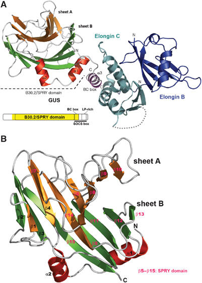
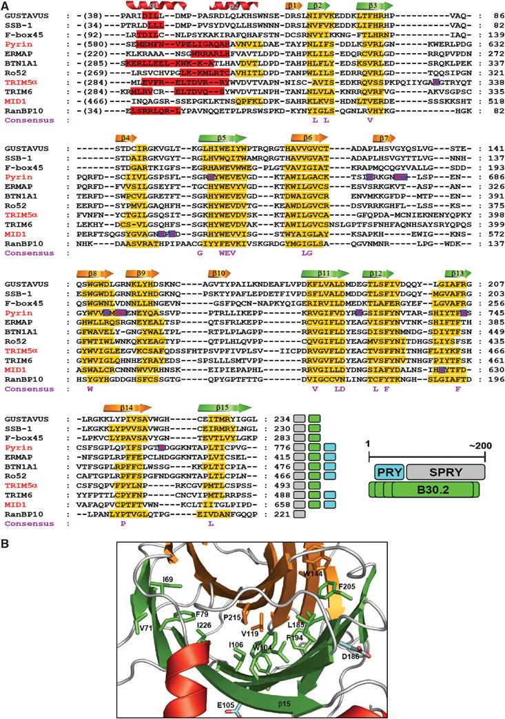
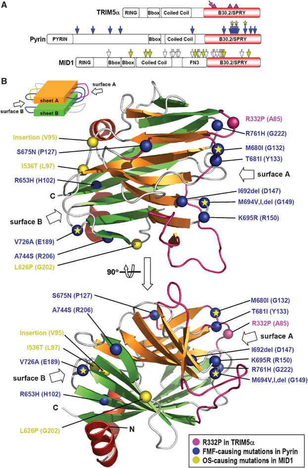
The B30.2/SPRY domain is present in approximately 700 eukaryotic (approximately 150 human) proteins, including medically important proteins such as TRIM5alpha and Pyrin. Nonetheless, the functional role of this modular domain remained unclear. Here, we report the crystal structure of an SPRY-SOCS box family protein GUSTAVUS in complex with Elongins B and C, revealing a highly distorted two-layered beta-sandwich core structure of its B30.2/SPRY domain. Ensuing studies identified one end of the beta-sandwich as the surface interacting with an RNA helicase VASA with a 40 nM dissociation constant. The sequence variation in TRIM5alpha responsible for HIV-1 restriction and most of the mutations in Pyrin causing familial Mediterranean fever map on this surface, implicating the corresponding region in many B30.2/SPRY domains as the ligand-binding site. The amino acids lining the binding surface are highly variable among the B30.2/SPRY domains, suggesting that these domains are protein-interacting modules, which recognize a specific individual partner protein rather than a consensus sequence motif.


Figures

Figure 1
Figure 1
Structure of the GUS:ElonginBC complex. (A) Ribbon drawing of the GUS:ElonginBC complex. The α-helices of GUS are in either red or magenta (for BC box), sheet A in orange, and sheet B in green. The domain organization of GUSTAVUS is shown. The region in yellow on the diagram indicates the fragment of the protein used for the structure determination. (B) Ribbon drawing of the B30.2/SPRY domain of GUS. The secondary structural elements are sequentially labeled and colored as in (A). The SPRY domain in GUS is indicated by color-coding the secondary structural elements.
Figure 2
Figure 2
Multiple sequence alignment and consensus residues. (A) Sequence alignment and secondary structure prediction of B30.2/SPRY domains. In total, 20 sequences of SPRY domains and ∼60 preceding amino acids were aligned (Supplementary Figure 3), and 11 of these are shown. The sequences were first aligned using ClustalX (Thompson et al, 1997) for a global alignment, followed by manual adjustments based on the consensus secondary structure prediction using NPSA (Deleage et al, 1997) and Jpred (Cuff et al, 1998). The secondary structures predicted by Jpred are indicated by red bars for α-helices and yellow bars for β-strands. The crystallographically determined secondary structural elements of GUS are shown at the top of the alignment. The letters at the bottom of the alignment indicate the similarity consensus residues conserved in the 20 sequences. The red letters in blue box indicate the positions of the mutations causing FMF (in pyrin), OS (in MID1), or the single amino-acid substitution in human TRIM5α that confers the ability to restrict HIV-1. The accession numbers for the sequences are listed in the Materials and methods section. The colored boxes at the end of each sequence indicate the domains identified in the databases: gray, SPRY domain; green, B30.2 domain; cyan, PRY domain. The relationship between the three domains on the primary structure is schematically drawn. Varying length of the B30.2 domains is indicated by multiple lines. (B) Location of the consensus residues. The consensus residues (shown in A) are represented as sticks on the ribbon drawing of the B30.2/SPRY domain of GUS and most of them are labeled. The hydrophobic residues on sheets A and B are in orange and green, respectively, while the hydrophilic residues are in cyan.
Figure 3
Figure 3
Sequence variations or mutations in TRIM5α, Pyrin, and MID1. (A) Location on the primary structures. The diagrams depict the primary structures of the three proteins and the domains they possess. The locations of the sequence variations or disease-causing mutations are marked on the diagrams for TRIM5α: pink triangles, locations of the significant sequence variation and length polymorphism in primate TRIM5α proteins; pink arrow, the substitution of R332P in human TRIM5α that confers the ability to restrict HIV-1, for Pyrin: blue arrows, the FMF-causing point mutations including three mutational hot spots marked with an asterisk, and for MID1: white arrows, the OS-causing frame shift or nonsense mutations; yellow arrows, point mutations, insertion, or deletion of amino acids. (B) Location of the corresponding residues of GUS on the tertiary structure. The sequence variations or mutations in the three proteins are mapped on the structure of the B30.2/SPRY domain of GUS. The mutation sites are indicated by large Cα atom spheres and labels shown in the same color of the arrows in (A). The residues of GUS corresponding to the mutation points are in the parentheses. The loop regions in pink correspond to the locations of the length polymorphism in the primate TRIM5α proteins. ‘Insertion' stands for the eight amino-acid insertional mutation in MID1, and ‘del' stands for deletion of a residue in Pyrin. A schematic drawing of the β-sandwich structure of GUS is shown to aid the recognition of surface A and surface B.
Figure 4
Figure 4
Identification of the GUS-binding region of VASA. (A) (His)6-tag pull-down experiment. Each of the indicated (His)6-tagged MBP-fused VASA deletion mutants and GUS without a tag were mixed and incubated with Ni-NTA agarose resin (QIAGEN). The resin was vigorously washed and subjected to denaturing gel electrophoresis. (B) ITC analysis of the interaction between GUS and VASA. The ITC experiments were carried out by titrating 5 μl of 0.15 mM of GUS into the solution of full-length VASA (5 μM) or titrating 5 μl of 0.2 mM of the 30 amino-acid VASA peptide into the solution of GUS (9 μM) or GUS in complex with ElonginBC (9 μM). KD values were deduced from curve fittings of the integrated heat per mol of added ligand and presented in the table. The titration curves obtained for the formation of the indicated complexes are shown in the insets.
Figure 5
Figure 5
Identification of the VASA-binding surface of GUS. (A) Locations of the site-directed mutagenesis on the B30.2/SPRY domain of GUS. The green dotted circles and labels on surface A indicate the mutations that decreased the binding affinity for the VASA peptide. The dotted circles in magenta indicate the mutations on surface B, which did not affect the binding affinity. The residues of GUS corresponding to the sequence variation and the disease-related mutations in TRIM5α, Pyrin, and MID1 are highlighted by the indicated colors. (B) ITC analysis of the interaction between GUS mutants and the VASA peptide. The titration curves and curve fittings are shown for two indicated GUS mutants and the KD values determined for the three GUS mutants are tabulated. (C) Denaturing gel elelctrophoretic analysis of the interaction between GUS mutants and the VASA peptide. Wild-type GUS and the mutants in complex with ElonginBC (each at 10 μM) were mixed with VASA peptide (30 μM) and incubated for 2 h. The mixtures were dialyzed for one day at 4°C, and each sample in the dialysis bags was subjected to denaturing gel electrophoresis. The mutants containing the amino-acid substitution on surface A or surface B are labeled with green and magenta letters, respectively. (D) Native gel electrophoretic analysis of the interaction between GUS mutants and full-length VASA. Wild-type and mutant GUS proteins in complex with ElonginBC (each at 10 μM) were mixed with VASA (5 μM) and incubated for 4 h. The analysis of the reaction mixtures by gel electrophoresis is shown. The amino-acid substitutions in the mutant proteins are color-coded as in C. While VASA mixed with wild-type GUS migrates to a distinct location, signifying a complex formation between the two, neither reduction of the band intensity nor appearance of a new band is observable for VASA mixed with the indicated GUS mutants.

References

    1. Bakkaloglu A (2003) Familial Mediterranean fever. Pediatr Nephrol 18: 853–859 - PubMed
    1. Bateman A, Coin L, Durbin R, Finn RD, Hollich V, Griffiths-Jones S, Khanna A, Marshall M, Moxon S, Sonnhammer EL, Studholme DJ, Yeats C, Eddy SR (2004) The Pfam protein families database. Nucleic Acids Res 32: D138–D141 - PMC - PubMed
    1. Brunger AT, Adams PD, Clore GM, DeLano WL, Gros P, Grosse-Kunstleve RW, Jiang JS, Kuszewski J, Nilges M, Pannu NS, Read RJ, Rice LM, Simonson T, Warren GL (1998) Crystallography & NMR system: A new software suite for macromolecular structure determination. Acta Crystallogr D Biol Crystallogr 54: 905–921 - PubMed
    1. Carrera P, Johnstone O, Nakamura A, Casanova J, Jackle H, Lasko P (2000) VASA mediates translation through interaction with a Drosophila yIF2 homolog. Mol Cell 5: 181–187 - PubMed
    1. Cuff JA, Clamp ME, Siddiqui AS, Finlay M, Barton GJ (1998) JPred: a consensus secondary structure prediction server. Bioinformatics 14: 892–893 - PubMed

Publication types

MeSH terms

Associated data