. 2006 Mar;116(3):825-32.
doi: 10.1172/JCI27120. Epub 2006 Feb 23.

Cholinergic dysfunction in a mouse model of Alzheimer disease is reversed by an anti-A beta antibody

Affiliations

Cholinergic dysfunction in a mouse model of Alzheimer disease is reversed by an anti-A beta antibody

Kelly R Bales et al. J Clin Invest. 2006 Mar.

Abstract

Disruption of cholinergic neurotransmission contributes to the memory impairment that characterizes Alzheimer disease (AD). Since the amyloid cascade hypothesis of AD pathogenesis postulates that amyloid beta (A beta) peptide accumulation in critical brain regions also contributes to memory impairment, we assessed cholinergic function in transgenic mice where the human A beta peptide is overexpressed. We first measured hippocampal acetylcholine (ACh) release in young, freely moving PDAPP mice, a well-characterized transgenic mouse model of AD, and found marked A beta-dependent alterations in both basal and evoked ACh release compared with WT controls. We also found that A beta could directly interact with the high-affinity choline transporter which may impair steady-state and on-demand ACh release. Treatment of PDAPP mice with the anti-A beta antibody m266 rapidly and completely restored hippocampal ACh release and high-affinity choline uptake while greatly reducing impaired habituation learning that is characteristic of these mice. Thus, soluble "cholinotoxic" species of the A beta peptide can directly impair cholinergic neurotransmission in PDAPP mice leading to memory impairment in the absence of overt neurodegeneration. Treatment with certain anti-A beta antibodies may therefore rapidly reverse this cholinergic dysfunction and relieve memory deficits associated with early AD.


Figures

Figure 1
Figure 1
Hippocampal ACh release and tissue levels are reduced in PDAPP transgenic mice. (A) Basal levels of hippocampal ACh release measured by in vivo microdialysis from WT and PDAPP transgenic mice (n = 7–10 mice per group, 4–6 months of age). (B and C) Tissue levels of ACh from the hippocampus (B) and cortex (C) of PDAPP and WT mice (n = 6 mice per group). *P < 0.05 versus WT.
Figure 2
Figure 2
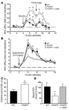
Hippocampal ACh release in PDAPP mice is restored to WT levels after anti-Aβ treatment. (A) Hippocampal ACh release in WT and PDAPP transgenic mice following exposure to a novel environment (Novelty) and after treatment of PDAPP mice with the anti-Aβ antibody m266 (PDAPP + m266; 500 μg i.p.). The arrows indicate the time at which mice were placed into a novel environment (novelty) and back into their home cage. (B) Release of ACh from the hippocampus of WT and PDAPP mice following administration of scopolamine (0.3 mg/kg i.p.) and after administration of m266. n = 7–10 mice per group, 4–6 months of age. (C) Extracellular levels of choline in brain measured by in vivo microdialysis from WT and PDAPP mice. n = 5 mice per group, 4–6 months of age. (D) Basal levels of hippocampal ACh release in WT, PDAPP, and PDAPP mice administered m266. n = 7–10 mice per group, 4–6 months of age. *P < 0.05 versus WT; #P < 0.05, PDAPP versus PDAPP + m266.
Figure 3
Figure 3
Aβ interacts with ChT-1. (A and B) High-affinity choline uptake into rat synaptosomes (A) and a cell line expressing the human ChT-1 (B) was significantly increased after exposure to Aβ42. Data are the percent of choline uptake in untreated, control samples (mean ± SEM of triplicate values) from 1 representative experiment, which was repeated 2–3 times with similar results. *P < 0.05. (C) ChT-1 was coimmunoprecipitated from hippocampal extracts by the anti-Aβ antibody 4G8 followed by Western blot analysis with an anti–ChT-1 antibody. (D) The Aβ peptide was coimmunoprecipitated from hippocampal extracts prepared from PDAPP transgenic mice by an anti–ChT-1 antibody followed by Western blot analysis with biotinylated anti-Aβ antibodies 21F12 and 2G3. (E) The anti-Aβ42 antibody 21F12, but not the anti-Aβ40 antibody 2G3, coimmunoprecipitated ChT-1 from hippocampal extracts prepared from PDAPP transgenic mice followed by Western blot analysis with an anti–ChT-1 antibody. (F) Neither an irrelevant IgG nor an anti–glutamate 1 transporter antibody (EAAT-1) coimmunoprecipitated Aβ from hippocampal extracts prepared from PDAPP mice, whereas Aβ was readily detected following immunoprecipitation with an antibody directed toward ChT-1 followed by Western blot analysis with an anti-Aβ antibody.
Figure 4
Figure 4
Relative mRNA level of ChT-1 in the hippocampus of young (2 months of age) and old (17 months of age) WT and PDAPP transgenic mice as determined by real-time quantitative PCR amplification. n = 5 mice per group. **P < 0.01, *P < 0.05 versus respective 17-month-old groups.
Figure 5
Figure 5
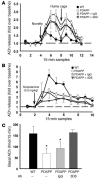
Hippocampal ACh release is unaltered after treatment with other IgGs. (A) Hippocampal release of ACh in WT and PDAPP transgenic mice following exposure to a novel environment and 24 hours after the administration of an irrelevant IgG (PDAPP + IgG; 500 μg i.p.) or the anti-Aβ antibody 3D6 (PDAPP + 3D6; 500 μg i.p.). (B) Release of ACh from the hippocampus of PDAPP and WT mice following administration of scopolamine (0.3 mg/kg i.p.) 24 hours after the administration of an irrelevant IgG or the anti-Aβ antibody 3D6. No significant difference between treated PDAPP and untreated PDAPP mice were observed in either A or B. (C) Basal levels of hippocampal ACh release from WT and PDAPP mice 24 hours following administration of an irrelevant IgG or the anti-Aβ antibody 3D6. *P < 0.05 versus WT and PDAPP + 3D6. n = 3–10 mice per group, 4–6 months of age.
Figure 6
Figure 6
Habituation learning in a novel open field in PDAPP mice is normalized after treatment with m266. (A) PDAPP transgenic mice exhibited increased locomotor activity, which was reduced following treatment with the anti-Aβ antibody m266 (500 μg i.p.) but not following treatment with the anti-Aβ antibody 3D6 (500 μg i.p.). (B) Accumulated locomotor activity per 60-minute session. ***P < 0.001. n = 20 mice per group, 4–6 months of age.

References

    1. Perry E, Walker M, Grace J, Perry R. Acetylcholine in mind: a neurotransmitter correlate of consciousness? Trends Neurosci. 1999;22:273–280. - PubMed
    1. Bartus R, Dean RL, Beer B, Lippa AS. The cholinergic hypothesis of geriatric memory dysfunction. Science. 1982;217:408–411. - PubMed
    1. Coyle JT, Price DL, DeLong MR. Alzheimer’s disease: a disorder of cortical cholinergic innervation. Science. 1983;219:1184–1190. - PubMed
    1. Whitehouse PJ, et al. Alzheimer disease: evidence for selective loss of cholinergic neurons in the nucleus basalis. Ann. Neurol. 1981;10:122–126. - PubMed
    1. Hardy J, Selkoe DJ. The amyloid hypothesis of Alzheimer’s disease: progress and problems on the road to therapeutics. Science. 2002;297:353–356. - PubMed

MeSH terms