

Intranasal administration of the growth-compromised HSV-2 vector DeltaRR prevents kainate-induced seizures and neuronal loss in rats and mice
- PMID: 16500153
- PMCID: PMC1513123
- DOI: 10.1016/j.ymthe.2005.12.013
Intranasal administration of the growth-compromised HSV-2 vector DeltaRR prevents kainate-induced seizures and neuronal loss in rats and mice
Erratum in
- Mol Ther. 2007 Sep;15(9):1734
Abstract
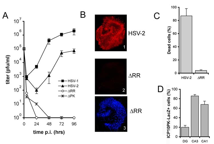
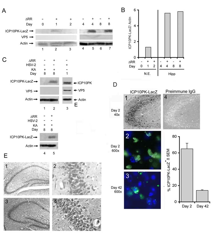
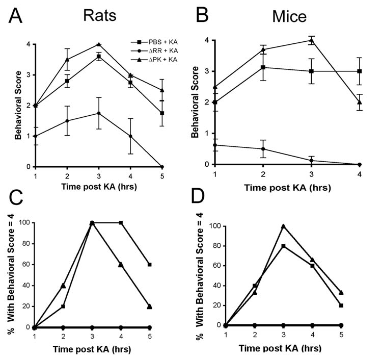
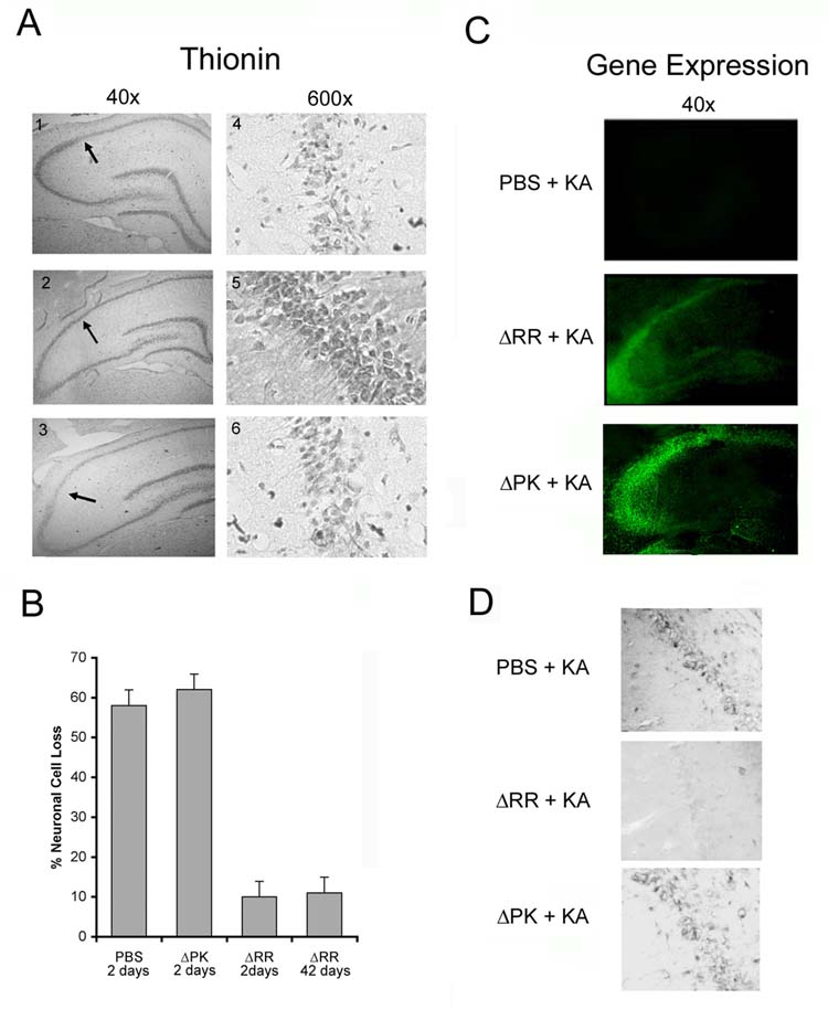
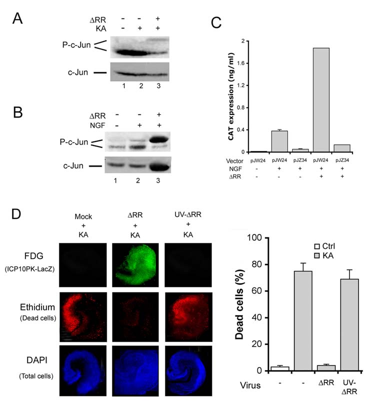
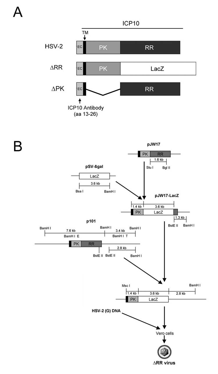
Identification of targets and delivery platforms for gene therapy of neurodegenerative disorders is a clinical challenge. We describe a novel paradigm in which the neuroprotective gene is the herpes simplex virus type 2 (HSV-2) antiapoptotic gene ICP10PK and the vector is the growth-compromised HSV-2 mutant DeltaRR. DeltaRR is delivered intranasally. It is not toxic in rats and mice. ICP10PK is expressed in the hippocampus of the DeltaRR-treated animals for at least 42 days in the absence of virus replication and late virus gene expression. Its expression is regulated by an AP-1 amplification loop. Intranasally delivered DeltaRR prevents kainic acid-induced seizures, neuronal loss, and inflammation, in both rats and mice. The data suggest that DeltaRR is a promising therapeutic platform for neurodegenerative diseases.
Figures

Similar articles
-
The growth compromised HSV-2 mutant DeltaRR prevents kainic acid-induced apoptosis and loss of function in organotypic hippocampal cultures.Brain Res. 2006 Nov 13;1119(1):26-39. doi: 10.1016/j.brainres.2006.08.078. Epub 2006 Oct 3. Brain Res. 2006. PMID: 17020750 Free PMC article.
-
Multi-targeted neuroprotection by the HSV-2 gene ICP10PK includes robust bystander activity through PI3-K/Akt and/or MEK/ERK-dependent neuronal release of vascular endothelial growth factor and fractalkine.J Neurochem. 2010 Feb;112(3):662-76. doi: 10.1111/j.1471-4159.2009.06475.x. Epub 2009 Nov 5. J Neurochem. 2010. PMID: 19891735 Free PMC article.
-
Growth-compromised HSV-2 vector Delta RR protects from N-methyl-D-aspartate-induced neuronal degeneration through redundant activation of the MEK/ERK and PI3-K/Akt survival pathways, either one of which overrides apoptotic cascades.J Neurosci Res. 2008 Feb 1;86(2):378-91. doi: 10.1002/jnr.21486. J Neurosci Res. 2008. PMID: 17893911
-
The HSV-2 protein ICP10PK prevents neuronal apoptosis and loss of function in an in vivo model of neurodegeneration associated with glutamate excitotoxicity.Exp Neurol. 2007 Feb;203(2):381-93. doi: 10.1016/j.expneurol.2006.08.022. Epub 2006 Oct 16. Exp Neurol. 2007. PMID: 17046754 Free PMC article.
-
Herpes simplex virus vectors overexpressing the glucose transporter gene protect against seizure-induced neuron loss.Proc Natl Acad Sci U S A. 1995 Aug 1;92(16):7247-51. doi: 10.1073/pnas.92.16.7247. Proc Natl Acad Sci U S A. 1995. PMID: 7638175 Free PMC article.
Cited by
-
Calpain-dependent clearance of the autophagy protein p62/SQSTM1 is a contributor to ΔPK oncolytic activity in melanoma.Gene Ther. 2014 Apr;21(4):371-8. doi: 10.1038/gt.2014.6. Epub 2014 Feb 20. Gene Ther. 2014. PMID: 24553345 Free PMC article.
-
Phage-mimicking antibacterial core-shell nanoparticles.Nanoscale Adv. 2019 Nov 7;1(12):4812-4826. doi: 10.1039/c9na00461k. eCollection 2019 Dec 3. Nanoscale Adv. 2019. PMID: 36133139 Free PMC article.
-
Gene transfer to the outflow tract.Exp Eye Res. 2017 May;158:73-84. doi: 10.1016/j.exer.2016.04.023. Epub 2016 Apr 27. Exp Eye Res. 2017. PMID: 27131906 Free PMC article. Review.
-
Gene therapy for epilepsy.Metab Brain Dis. 2010 Sep;25(3):363-6. doi: 10.1007/s11011-010-9209-7. Epub 2010 Sep 23. Metab Brain Dis. 2010. PMID: 20862604 Review. No abstract available.
-
Sequestering survivin to functionalized nanoparticles: a strategy to enhance apoptosis in cancer cells.Biomater Sci. 2016 Apr;4(4):614-26. doi: 10.1039/c5bm00580a. Epub 2016 Feb 4. Biomater Sci. 2016. PMID: 26845086 Free PMC article.
References
-
- Natsume A, et al. Bcl-2 and GDNF delivered by HSV-mediated gene transfer act additively to protect dopaminergic neurons from 6-OHDA-induced degeneration. Exp. Neurol. 2001;169:231–238. - PubMed
-
- Kalwy SA, Akbar MT, Coffin RS, de Belleroche J, Latchman DS. Heat shock protein 27 delivered via a herpes simplex virus vector can protect neurons of the hippocampus against kainic-acid-induced cell loss. Brain Res. Mol. Brain Res. 2003;111:91–103. - PubMed
-
- Najioullah F, et al. Diagnosis and surveillance of herpes simplex virus infection of the central nervous system. J. Med. Virol. 2000;61:468–73. - PubMed
-
- Perkins D, Gyure KA, Pereira EF, Aurelian L. Herpes simplex virus type 1-induced encephalitis has an apoptotic component associated with activation of c-Jun N-terminal kinase. J. Neurovirol. 2003;9:101–111. - PubMed
-
- Perkins D, Pereira EF, Aurelian L. The herpes simplex virus type 2 R1 protein kinase (ICP10 PK) functions as a dominant regulator of apoptosis in hippocampal neurons involving activation of the ERK survival pathway and upregulation of the antiapoptotic protein Bag-1. J. Virol. 2003;77:1292–1305. - PMC - PubMed
Publication types
MeSH terms
Substances
Grants and funding
LinkOut - more resources
Full Text Sources
Other Literature Sources
Medical
