Review
. 2006 Mar;44(2):295-315, ix.
doi: 10.1016/j.rcl.2005.10.009.

Acute lung infections in normal and immunocompromised hosts

Affiliations
Review

Acute lung infections in normal and immunocompromised hosts

Stephen Waite et al. Radiol Clin North Am. 2006 Mar.

Abstract

Pulmonary infections are among the most common causes of morbidity and mortality worldwide, and contribute substantially to annual medical expenditures in the United States. Despite the availability of antimicrobial agents, pneumonia constitutes the sixth most common cause of death and the number one cause of death from infection. Pneumonia can be particularly life-threatening in the elderly, in individuals who have pre-existing heart and lung conditions, in patients who have suppressed or weakened immunity, and in pregnant women. This article discusses some of the important causes of acute lung infections in normal and immunocompromised hosts. Because there often is considerable overlap, infections are categorized by the host immune status that is most likely to be associated with a particular pathogen.


Figures

Fig. 1
Fig. 1
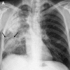
Streptococcus pneumonia. Chest radiograph demonstrates classic lobar pneumonia in the right upper lobe. Air space disease is homogeneous and confluent and there is no evidence of volume loss.
Fig. 2
Fig. 2
Streptococcus pneumonia. CT scan of the chest in another patient demonstrates a classic appearance of lobar pneumonia with confluent homogenous opacification of the right middle lobe. Air-bronchograms are noted (arrow).
Fig. 3
Fig. 3
Staphylococcus aureus pneumonia. Chest radiograph demonstrates multifocal opacities (arrows) predominantly in the left lung.
Fig. 4
Fig. 4
Staphylococcus aureus bronchopneumonia. CT scan of the chest in another patient demonstrates bilateral multifocal opacities. Bronchopneumonia often is associated with pleural effusions as seen in this patient.
Fig. 5
Fig. 5
Staphylococcus pneumonia. CT scan of the chest demonstrates multifocal inhomogeneous opacities consistent with bronchopneumonia (arrows) in addition to bilateral pleural effusions and secondary relaxation atelectasis.
Fig. 6
Fig. 6
Staphylococcus pneumonia. CT scan of the chest through the upper lobes of the same patient as in Fig. 5 demonstrates dense consolidation in the left upper lobe containing a focal region of low attenuation and gas (arrow) that is consistent with necrosis and abscess formation.
Fig. 7
Fig. 7
Mycoplasma pneumoniae. CT scan of the chest demonstrates foci of ground glass attenuation (arrow, A). More inferiorly, CT scan of the chest demonstrates centrilobular nodules in a “tree-in-bud” pattern (circles, A and B).
Fig. 7
Fig. 7
Mycoplasma pneumoniae. CT scan of the chest demonstrates foci of ground glass attenuation (arrow, A). More inferiorly, CT scan of the chest demonstrates centrilobular nodules in a “tree-in-bud” pattern (circles, A and B).
Fig. 8
Fig. 8
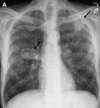
Legionella pneumonia in a patient who presented with severe respiratory distress. (A) Chest radiograph demonstrates multifocal bilateral air space disease. (B) Within 2 weeks, the patient's respiratory status continued to decline and he developed a left-sided pneumothorax.
Fig. 8
Fig. 8
Legionella pneumonia in a patient who presented with severe respiratory distress. (A) Chest radiograph demonstrates multifocal bilateral air space disease. (B) Within 2 weeks, the patient's respiratory status continued to decline and he developed a left-sided pneumothorax.
Fig. 9
Fig. 9
Aspiration pneumonia in a 65-year-old alcoholic who had respiratory distress. (A) Admission chest radiograph demonstrates a right pleural effusion and multiple cavities containing air–fluid levels in the medial aspect of the right middle lung zone (arrows). (B) Lateral view localizes the cavities to the superior segment of the right lower lobe (arrow). (C) CT scan of the chest demonstrates multiple cavities in a dependent location in the medial aspect of the superior segment of the right upper lobe. (D) Images through the lung bases demonstrate right middle lobe consolidation and nondependent parapneumonic effusion.
Fig. 9
Fig. 9
Aspiration pneumonia in a 65-year-old alcoholic who had respiratory distress. (A) Admission chest radiograph demonstrates a right pleural effusion and multiple cavities containing air–fluid levels in the medial aspect of the right middle lung zone (arrows). (B) Lateral view localizes the cavities to the superior segment of the right lower lobe (arrow). (C) CT scan of the chest demonstrates multiple cavities in a dependent location in the medial aspect of the superior segment of the right upper lobe. (D) Images through the lung bases demonstrate right middle lobe consolidation and nondependent parapneumonic effusion.
Fig. 10
Fig. 10
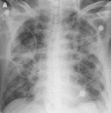
Viral pneumonia. CXR demonstrates a diffuse bilateral reticular pattern consistent with viral infection. Note the presence of Kerley lines (circle), an uncommon feature in viral pneumonia.
Fig. 11
Fig. 11
Influenzae pneumonia. CT scan of the chest demonstrates patchy multifocal ground glass attenuation opacities (arrows).
Fig. 12
Fig. 12
Influenzae pneumonia. CT scan of another patient who has influenza pneumonia demonstrates areas of bronchial wall thickening (arrow) and airway disease.
Fig. 13
Fig. 13
Fibrosing mediastinitis in a 54-year-old man who had chronic histoplasmosis. CT scan of the chest demonstrates calcified mediastinal and hilar adenopathy. Note the narrowing of the left main pulmonary artery. The right main pulmonary artery is truncated and no right pulmonary veins are patent on imaging (incompletely seen).
Fig. 14
Fig. 14
Coccidiodes immitis was isolated at bronchoscopy in a 34-year-old Mexican patient who presented to the emergency department with mild respiratory symptoms. (A and B) CT scan at two levels through the bases demonstrates multiple nodular areas of consolidation in the left lower lobe.
Fig. 14
Fig. 14
Coccidiodes immitis was isolated at bronchoscopy in a 34-year-old Mexican patient who presented to the emergency department with mild respiratory symptoms. (A and B) CT scan at two levels through the bases demonstrates multiple nodular areas of consolidation in the left lower lobe.
Fig. 15
Fig. 15
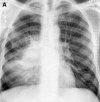
PCP pneumonia in a young HIV-positive patient. CXR demonstrates predominantly central air-space disease with peripheral sparing.
Fig. 16
Fig. 16
PCP pneumonia in another young HIV-positive patient. CT scan demonstrates a mixed pattern of ground glass attenuation and superimposed prominent septal lines in a “crazy-paving” pattern.
Fig. 17
Fig. 17
PCP pneumonia. CT scan of the chest demonstrates cystic air spaces of varying sizes that are consistent with pneumatoceles.
Fig. 18
Fig. 18
PCP pneumonia in an HIV-positive patient who had hypoxia. CT scan of the chest demonstrates an atypical pattern with scattered irregular heterogeneous densities and areas of bronchial wall thickening.
Fig. 19
Fig. 19
(A) Cryptococcus infection in an HIV-positive patient who had respiratory distress. CXR demonstrates multiple bilateral foci of consolidation (arrows), some of which appear nodular and cavitary. Emphysematous changes are identified at the apices, especially on the right. (B) Accompanying CT coronal image demonstrates upper lobe cystic air space disease. Bilateral upper lobe nodules, cavitary in the left upper lobe, are demonstrated.
Fig. 19
Fig. 19
(A) Cryptococcus infection in an HIV-positive patient who had respiratory distress. CXR demonstrates multiple bilateral foci of consolidation (arrows), some of which appear nodular and cavitary. Emphysematous changes are identified at the apices, especially on the right. (B) Accompanying CT coronal image demonstrates upper lobe cystic air space disease. Bilateral upper lobe nodules, cavitary in the left upper lobe, are demonstrated.
Fig. 20
Fig. 20
Primary TB. (A) CXR demonstrates prominent unilateral right hilar adenopathy. (B) CT scan demonstrates necrotic subcarinal and right hilar adenopathy.
Fig. 20
Fig. 20
Primary TB. (A) CXR demonstrates prominent unilateral right hilar adenopathy. (B) CT scan demonstrates necrotic subcarinal and right hilar adenopathy.
Fig. 21
Fig. 21
Postprimary TB in an immunocompromised patient who had weight loss and night sweats. (A) CXR demonstrates biapical cavitary consolidation. (B) CT scan confirms the cavitary nature of upper lobe opacities.
Fig. 21
Fig. 21
Postprimary TB in an immunocompromised patient who had weight loss and night sweats. (A) CXR demonstrates biapical cavitary consolidation. (B) CT scan confirms the cavitary nature of upper lobe opacities.
Fig. 22
Fig. 22
Miliary TB. Culture proven miliary TB in an HIV-positive patient with several weeks' duration of constitutional symptoms, fever, and weight loss. (A) CXR demonstrates biapical cavitary lesions and superimposed innumerable diffuse well-defined subcentimeter nodules. (B and C) CT scan confirms presence of upper lobe consolidation and innumerable randomly distributed subcentimeter nodules consistent with a miliary distribution. Some nodules (arrows, C) are on pleural surfaces, an important differentiation from airway nodules which are separate from the pleura.
Fig. 22
Fig. 22
Miliary TB. Culture proven miliary TB in an HIV-positive patient with several weeks' duration of constitutional symptoms, fever, and weight loss. (A) CXR demonstrates biapical cavitary lesions and superimposed innumerable diffuse well-defined subcentimeter nodules. (B and C) CT scan confirms presence of upper lobe consolidation and innumerable randomly distributed subcentimeter nodules consistent with a miliary distribution. Some nodules (arrows, C) are on pleural surfaces, an important differentiation from airway nodules which are separate from the pleura.
Fig. 23
Fig. 23
Mycobacterium avium–intracellularae pneumonia in a middle-aged man who had fever. (A) CXR demonstrates cavitary consolidation in the left upper lung zone (arrow). (B) Coronal CT confirms the presence of a thick-walled cavitary lesion in the left upper lobe. Imaging is indistinguishable from postprimary TB.
Fig. 23
Fig. 23
Mycobacterium avium–intracellularae pneumonia in a middle-aged man who had fever. (A) CXR demonstrates cavitary consolidation in the left upper lung zone (arrow). (B) Coronal CT confirms the presence of a thick-walled cavitary lesion in the left upper lobe. Imaging is indistinguishable from postprimary TB.
Fig. 24
Fig. 24
Mycobacterium avium–intracellularae pneumonia in a middle-aged woman. CT scan demonstrates evidence of small airway disease with scattered bilateral centrilobular “tree-in-bud” structures (circle). Bronchial wall thickening is seen in the right middle lobe.
Fig. 25
Fig. 25
Mycobacterium avium–intracellularae pneumonia in another patient. CT scan of the chest demonstrates bronchiectasis and bronchial wall thickening in the right middle lobe (arrow) and “tree-in-bud” structures in the lower lobes.
Fig. 26
Fig. 26
Acute aspergillus infection in a neutropenic patient. (A) CXR demonstrates large foci of nodular consolidation with “shaggy” borders in the right lung. (B) Corresponding CT scan confirms large nodular consolidation in the right upper lobe with surrounding heterogeneous ground glass attenuation. Findings are consistent with a “halo” sign.
Fig. 26
Fig. 26
Acute aspergillus infection in a neutropenic patient. (A) CXR demonstrates large foci of nodular consolidation with “shaggy” borders in the right lung. (B) Corresponding CT scan confirms large nodular consolidation in the right upper lobe with surrounding heterogeneous ground glass attenuation. Findings are consistent with a “halo” sign.
Fig. 27
Fig. 27
(A) CXR of the patient in Fig. 26 a couple of weeks later demonstrates that the previously identified nodules are better defined and have developed a peripheral crescent of air (arrows). (B) Corresponding CT scan demonstrates cavitary consolidation containing air-bronchograms and an “air crescent” sign (arrow). The “air crescent sign” results when air fills the space between devitalized tissue and surrounding parenchyma. In the appropriate clinical setting, this finding is specific for aspergillus infection.
Fig. 27
Fig. 27
(A) CXR of the patient in Fig. 26 a couple of weeks later demonstrates that the previously identified nodules are better defined and have developed a peripheral crescent of air (arrows). (B) Corresponding CT scan demonstrates cavitary consolidation containing air-bronchograms and an “air crescent” sign (arrow). The “air crescent sign” results when air fills the space between devitalized tissue and surrounding parenchyma. In the appropriate clinical setting, this finding is specific for aspergillus infection.
Fig. 28
Fig. 28
CMV pneumonia in a bone marrow transplant recipient. CT scan of the chest demonstrates ground glass attenuation and consolidation (arrow, A and B). Centrilobular air space nodule consistent with an airway distribution is identified in the lingula (circle, B). CMV was isolated at bronchoscopy.
Fig. 28
Fig. 28
CMV pneumonia in a bone marrow transplant recipient. CT scan of the chest demonstrates ground glass attenuation and consolidation (arrow, A and B). Centrilobular air space nodule consistent with an airway distribution is identified in the lingula (circle, B). CMV was isolated at bronchoscopy.
Fig. 29
Fig. 29
Anthrax. Blood culture confirmed case of inhalational anthrax in a 61-year-old postal worker who presented to the emergency room after 3 days of experiencing general malaise and chills. (A) Contrast-enhanced CT scan demonstrated diffuse mediastinal infiltration and large bilateral pleural effusions. Right pleural effusion has a fluid-fluid level with layering high attenuation fluid; consistent with hemorrhage (black arrows, A and B). (B and C) Delayed CT scan of the chest demonstrates high attenuation mediastinal and hilar adenopathy; consistent with hemorrhage (white arrow, C). Anthrax infection should be considered in cases with high-attenuation adenopathy without intravenous contrast administration. (Courtesy of Jeffrey Galvin, MD, Baltimore, MD and the Armed Forces Institute of Pathology).
Fig. 29
Fig. 29
Anthrax. Blood culture confirmed case of inhalational anthrax in a 61-year-old postal worker who presented to the emergency room after 3 days of experiencing general malaise and chills. (A) Contrast-enhanced CT scan demonstrated diffuse mediastinal infiltration and large bilateral pleural effusions. Right pleural effusion has a fluid-fluid level with layering high attenuation fluid; consistent with hemorrhage (black arrows, A and B). (B and C) Delayed CT scan of the chest demonstrates high attenuation mediastinal and hilar adenopathy; consistent with hemorrhage (white arrow, C). Anthrax infection should be considered in cases with high-attenuation adenopathy without intravenous contrast administration. (Courtesy of Jeffrey Galvin, MD, Baltimore, MD and the Armed Forces Institute of Pathology).
Fig. 30
Fig. 30
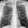
SARS in a 54-year-old Asian physician who was living in Toronto, Canada. (A) Initial radiograph demonstrates foci of consolidation in the left upper lobe. (B) Within 3 days there was marked progression with diffuse bilateral air space disease, and the patient required mechanical ventilation. A rapid progression is typical of SARS pneumonia. (Courtesy of Jeffrey Galvin, MD, Baltimore, MD and the Armed Forces Institute of Pathology).
Fig. 30
Fig. 30
SARS in a 54-year-old Asian physician who was living in Toronto, Canada. (A) Initial radiograph demonstrates foci of consolidation in the left upper lobe. (B) Within 3 days there was marked progression with diffuse bilateral air space disease, and the patient required mechanical ventilation. A rapid progression is typical of SARS pneumonia. (Courtesy of Jeffrey Galvin, MD, Baltimore, MD and the Armed Forces Institute of Pathology).

Similar articles

Cited by

References

    1. Reittner P., Ward S., Heyneman L. Pneumonia: high-resolution CT findings in 114 patients. Eur Radiol. 2003;13(3):515–521. - PubMed
    1. Webb W.R., Higgins C.B. Thoracic imaging-pulmonary and cardiovascular radiology. Lippincott Williams and Wilkens; Philadelphia: 2005.
    1. American Thoracic Society Guidelines for the initial management of adults with community acquired pneumonia: diagnosis, assessment of severity, and initial antimicrobial therapy. Am J Respir Crit Care Med. 2001;163:1730–1754. - PubMed
    1. National Center for Health Statistics National hospital discharge survey: annual summary 1990. Vital Health Stat. 1998;13:1–225. - PubMed
    1. Mandell L.A. Epidemiology and etiology of community acquired pneumonia. Infect Dis Clin North Am. 2004;18:761–776. - PMC - PubMed

MeSH terms