. 2006 Mar;85(3):564-72.
doi: 10.1016/j.fertnstert.2005.08.046.

Stromal cells from endometriotic lesions and endometrium from women with endometriosis have reduced decidualization capacity

Affiliations

Stromal cells from endometriotic lesions and endometrium from women with endometriosis have reduced decidualization capacity

Petra A B Klemmt et al. Fertil Steril. 2006 Mar.

Abstract

Objective: To evaluate the phenotype, proliferative, and differentiation capacities in vitro of stromal cells derived from peritoneal, ovarian, and deeply infiltrating endometriosis.

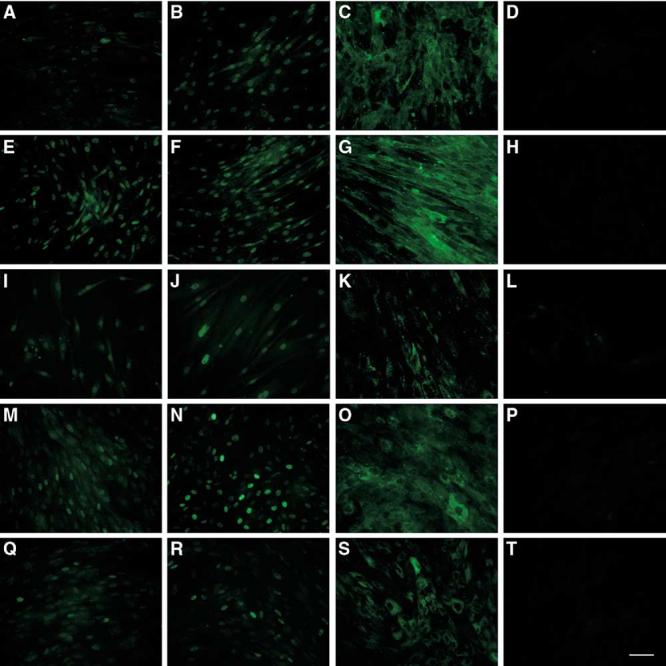
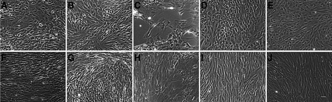
Design: Experimental study using phase contrast microscopy, immunocytochemistry, and functional bioassays.

Setting: University-based laboratory.

Patient(s): Women with and without endometriosis undergoing surgery for benign indications.

Intervention(s): None.

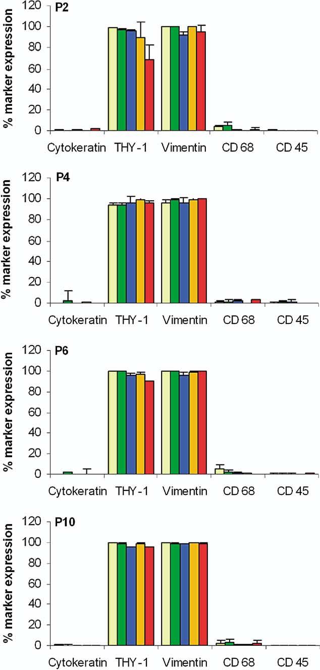
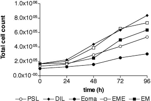
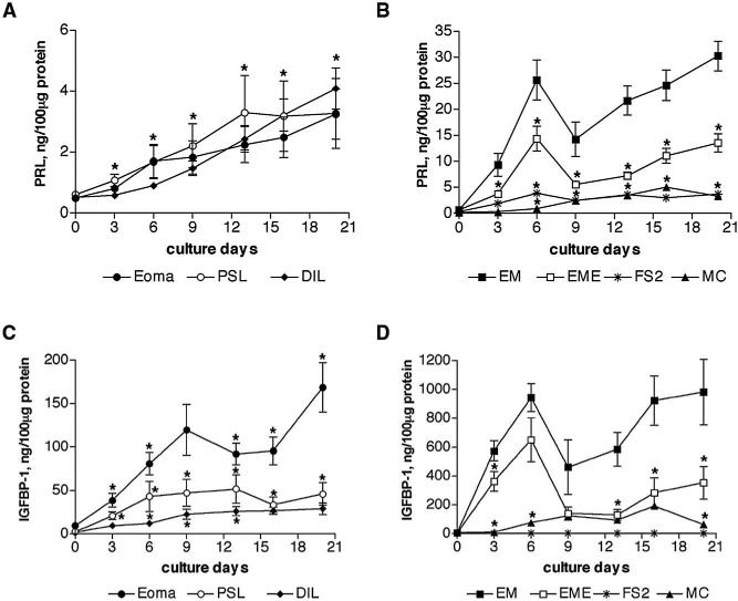
Main outcome measure(s): The stability in vitro of stromal cells derived from peritoneal (n = 18), ovarian (n = 29), and deeply infiltrating (n = 14) endometriotic lesions, as well as endometrium from women with (n = 5) and without endometriosis (n = 5) was evaluated by detection of endometrial markers. The proliferative and differentiation capacity of the cells was assessed by the use of cell doubling estimation and in vitro decidualization assays.

Result(s): The expression of the progesterone receptor and CD10 in stromal cells derived from the three types of endometriotic lesions is retained in culture up to passage 10. The doubling time of stromal cells from deeply infiltrating lesions is lower than that of endometrial stromal cells. Levels of prolactin and insulin-like growth factor binding protein-1 (IGFBP-1) are reduced in supernatants from stromal cells derived from the three types of lesions and from the endometrium of women with endometriosis.

Conclusion(s): The peritoneal, ovarian, and deeply infiltrating endometriotic stromal cell lines we describe retain in vivo tissue markers. Loss of differentiation capacity of the endometriotic cell lines and endometrial cells from women with endometriosis may influence the capacity for proliferation and survival of these cells in the ectopic environment.


Figures

FIGURE 1
FIGURE 1
Expression of markers in cultured stromal cells. Quantitative immunocytochemistry of tissue marker expression in endometriotic and endometrial stromal cells at passages 2, 4, 6, and 10. Data are expressed as percentage positive cells and bars represent mean ± SEM. Endometriotic stromal cells express >95% THY-1 and vimentin and <2% cytokeratin, CD45 and CD68. The level of purity and tissue marker expression is comparable to endometrial controls and remained stable with time in culture. (PSL, n = 8) peritoneal surface lesion; (DIL, n = 8) deeply infiltrating lesion; (Eoma, n = 8) ovarian endometrioma; (EME, n = 3) endometrium from women with endometriosis; (EM, n = 3) endometrium; and (P) passage of cells.
FIGURE 2
FIGURE 2
Expression of hormone receptors and CD10 in endometriotic and endometrial stromal cells. Endometriotic stromal cells (n = 3 each) from peritoneal surface lesions (AD), deeply infiltrating lesions (EH), ovarian endometrioma (IL), and endometrial stromal cells (n = 3 each) from women with (MP) and without endometriosis (QT) were grown on glass coverslips to confluence and stained for ER-α (A, E, I, M, Q), PR (B, F, J, N, R), and CD10 (C, G, K, O, S) expression and with the negative control mouse IgG (D, H, L, P, T) using immunofluorescent technique. The PSL and DIL stromal cell expressed ER-α at each passage tested, whereas Eoma stromal cells expressed ER-α only at primary culture. Expression of PR was detectable in all three endometriotic lesions at each passage tested. Endometrial stromal cells expressed both ER-α and PR. Endometriotic and endometrial stromal cells expressed CD10 in the cytoplasm. PSL, peritoneal surface lesion; DIL, deeply infiltrating lesion; Eoma, ovarian endometrioma. Magnification ×20, scale bar 100 μm.
FIGURE 3
FIGURE 3
Growth curve of endometriotic and endometrial stromal cells. Endometriotic and endometrial stromal cells were plated in DMEM complete, and total cell counts were determined after 24, 48, 72, and 96 hours of culture. (open circle, PSL, n = 3) peritoneal surface lesion; (diamond, DIL, n = 3) deeply infiltrating lesion; (filled circle, Eoma, n = 3) ovarian endometrioma; (open square, EME, n = 3) endometrium from women with endometriosis; (filled square, EM, n = 4) endometrium.
FIGURE 4
FIGURE 4
Morphology of in vitro decidualized endometriotic and endometrial stromal cells. Confluent stromal cells from peritoneal surface lesions (A, F), deeply infiltrating lesions (B, G), ovarian endometrioma (C, H), and endometrial stromal cells from women with (D, I) and without endometriosis (E, J) were treated with or without 0.5 mM of 8 Br-cAMP for 9 days. Endometriotic stromal cells (AC) underwent morphological changes from bipolar fibroblasts into polygonal decidual cells, but these were delayed (day 6 onward) and not as widespread compared with endometrial stromal cells that exhibited the characteristic change in morphology from day 3 onward (D, E). Untreated cells (FJ) retained a fibroblast-like, spindle shape appearance. Magnification ×10, scale bar 100 μm.
FIGURE 5
FIGURE 5
Time-dependent secretion of PRL and IGFBP-1 in endometriotic and endometrial stromal cells. Endometriotic stromal cells (A, C) and endometrial stromal cells (B, D) were allowed to decidualize in vitro, in response to 0.5 mM of 8-Br-cAMP for 20 days. The PRL and IGFBP-1 secretion by stromal cells into supernatants was measured every 3–4 days and normalized to total protein contents. Secretion of PRL was similar by all three types of endometriotic stromal cells, dermal fibroblasts, and myometrial myocytes. Endometrial stromal cells secreted tenfold more PRL in comparison with endometriotic stromal cells (*P<.001 throughout culture period). The PRL secretion by endometrial stromal cells from women with endometriosis was also reduced by half in comparison with normal endometrial stromal cells (*P<.05, B). (C) Secretion of IGFBP-1 was >threefold higher in stromal cells derived from ovarian endometrioma compared with peritoneal surface lesions and deeply infiltrating lesions. The difference between endometrioma and deeply infiltrating lesions was significant throughout the culture period. (D) Endometrial stromal cells secreted 20-fold more IGFBP-1 in comparison with endometriotic stromal cells (*P<.001 throughout the culture period). Secretion of IGFBP-1 was also lower in endometrial stromal cells from women with endometriosis compared with those from women without endometriosis (*P<.05). (○, PSL, n = 5) peritoneal surface lesion; (diamond, DIL, n = 5) deeply infiltrating lesion; (filled circle, Eoma, n = 5) ovarian endometrioma; (asterisk, FS2, n = 1) foreskin fibroblasts; (closed square, EM, n = 5) endometrium; (open square, EME, n = 5) endometrium from women with endometriosis; (triangle, MC, n = 1) myometrial myocytes.

Similar articles

Cited by

References

    1. Sampson J. The development of the implantation theory for the origin of peritoneal endometriosis. Am J Obstet Gynecol. 1940;40:549–56.
    1. Fujii S. Secondary Müllerian system and endometriosis. Am J Obstet Gynecol. 1991;165:219–25. - PubMed
    1. Nisolle M, Donnez J. Peritoneal endometriosis, ovarian endometriosis, and adenomyotic nodules of the rectovaginal septum are three different entities. Fertil Steril. 1997;68:585–96. - PubMed
    1. Osteen KG, Hill GA, Hargrove JT, Gorstein F. Development of a method to isolate and culture highly purified populations of stromal and epithelial cells from human endometrial biopsy specimens. Fertil Steril. 1989;52:965–72. - PubMed
    1. Fernandez-Shaw S, Shorter SC, Naish CE, Barlow DH, Starkey PM. Isolation and purification of human endometrial stromal and glandular cells using immunomagnetic microspheres. Hum Reprod. 1992;7:156–61. - PubMed

Publication types

MeSH terms