. 2006 Feb 25;34(4):1205-15.
doi: 10.1093/nar/gkj521. Print 2006.

The gene encoding the fragile X RNA-binding protein is controlled by nuclear respiratory factor 2 and the CREB family of transcription factors

Affiliations

The gene encoding the fragile X RNA-binding protein is controlled by nuclear respiratory factor 2 and the CREB family of transcription factors

Karen T Smith et al. Nucleic Acids Res. .

Abstract

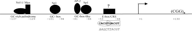
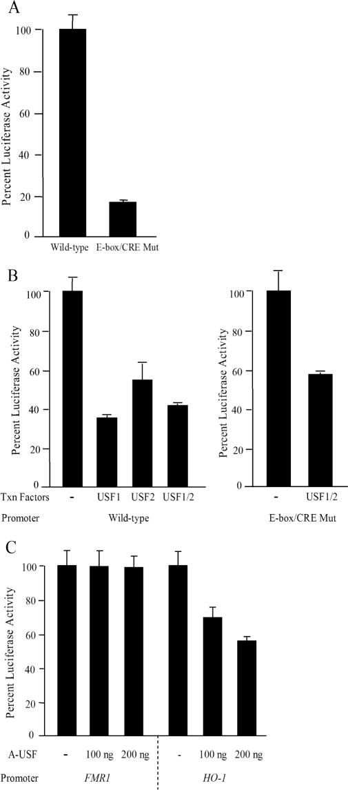
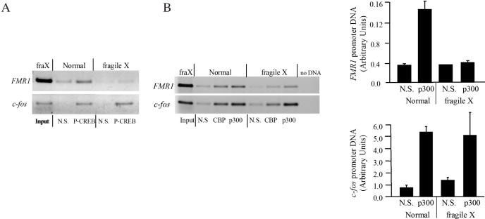
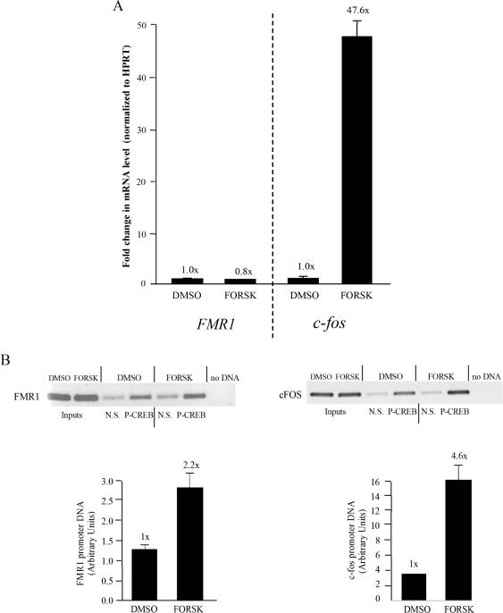
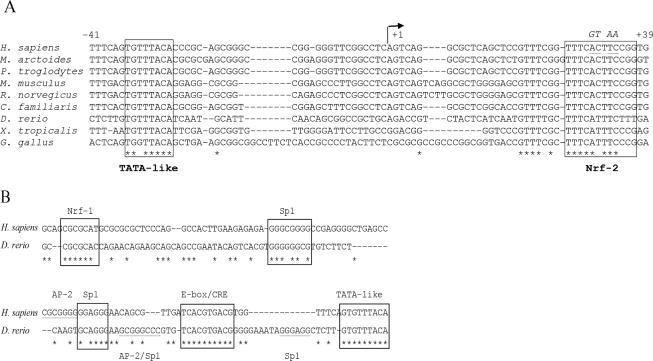
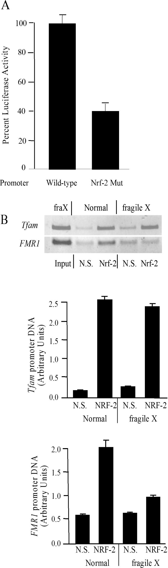
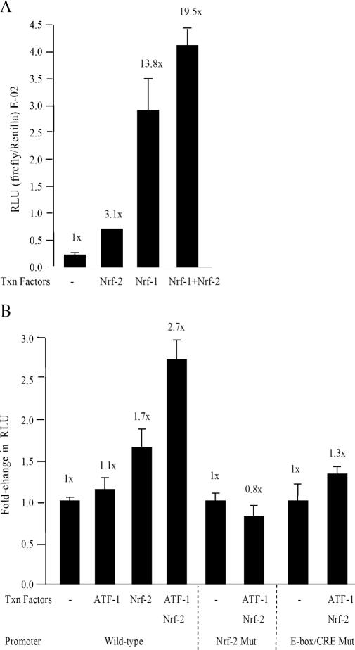
FMR1 encodes an RNA-binding protein whose absence results in fragile X mental retardation. In most patients, the FMR1 gene is cytosine-methylated and transcriptionally inactive. NRF-1 and Sp1 are known to bind and stimulate the active, but not the methylated/silenced, FMR1 promoter. Prior analysis has implicated a CRE site in regulation of FMR1 in neural cells but the role of this site is controversial. We now show that a phospho-CREB/ATF family member is bound to this site in vivo. We also find that the histone acetyltransferases CBP and p300 are associated with active FMR1 but are lost at the hypoacetylated fragile X allele. Surprisingly, FMR1 is not cAMP-inducible and resides in a newly recognized subclass of CREB-regulated genes. We have also elucidated a role for NRF-2 as a regulator of FMR1 in vivo through a previously unrecognized and highly conserved recognition site in FMR1. NRF-1 and NRF-2 act additively while NRF-2 synergizes with CREB/ATF at FMR1's promoter. These data add FMR1 to the collection of genes controlled by both NRF-1 and NRF-2 and disfavor its membership in the immediate early response group of genes.


Figures

Figure 1
Figure 1
Human FMR1 promoter map showing the four binding sites known to be occupied in vivo and the identities of the likely factors at those sites (12,13). The sequence of the overlapping E-box/CRE is shown underneath the site and that of the E-box/CRE mutant used in reporter assays is shown below the wild-type sequence with mutated bases underlined. A question mark above the E-box/CRE represents the unknown factor(s) binding this site in vivo. The bent arrow indicates the major transcription start site (61).
Figure 2
Figure 2
USF does not regulate transcription through the E-box. (A) HeLa cells were transfected with wild-type pFMR1-luc or with pFMR1-luc with point mutations in the E-box/CRE and luciferase activities were measured in cell lysates. (B) Either the wild-type (left) or E-box/CRE mutant reporter (right) was transfected into HeLa cells with or without pSV-USF1 and/or pSV-USF2 expression vectors as indicated. (C) pFMR1-luc or pHOGL3/4.5 was transfected into HeLa cells with the indicated amount of A-USF expression plasmid, or empty vector. Values for the FMR1 or HO-1 promoters were normalized to a CMV transfection control reporter. Data are expressed as percent normalized luciferase activity. Error bars represent ±1 standard deviation.
Figure 3
Figure 3
P-CREB/ATF and CBP/p300 occupy human FMR1 in vivo. (A) ChIP was carried out with either anti-P-CREB antibody or (B) antibodies to CBP and p300, or non-specific rabbit IgG [N.S. in both (A and B)]. PCR was performed to amplify the FMR1 and c-fos promoters in normal and fragile X cells (left). Occupancy of p300 was quantified by real-time PCR of an independent set of triplicate IPs (right); error bars represent ±1 standard deviation.
Figure 4
Figure 4
FMR1 transcription is not cAMP-inducible. (A) Real-time PCR quantification of endogenous FMR1, HPRT and c-fos transcripts in PC12 cells with or without 1 h of forskolin stimulation. (B) Phosphorylation of CREB/ATF at FMR1 increases 2-fold after forskolin induction of PC12 cells. ChIP was performed with P-CREB antiserum on DMSO- or forskolin-treated PC12 cells. PCR was performed to amplify the rat FMR1 and c-fos promoters (top). Quantification of P-CREB binding was performed by real-time PCR (bottom). Error bars represent ±1 standard deviation. For FMR1 real-time analysis, two independent IPs are represented, each with duplicate real-time PCR. For c-fos, the real-time data represent one IP with quadruplicate real-time PCR.
Figure 5
Figure 5
Identification and conservation of a NRF-2/GABP binding site in the 5′-UTR of FMR1. (A) Sequence alignment of FMR1 homologs from several species showing a newly identified binding site for NRF-2. GenBank accession nos are as follows: L29074.1 (Homo sapiens), AY630337 (Rattus norvegicus), AY630338 (Mus musculus), AF251348 (Canis familiaris), AF251350 (Macaca arctoides), AF251349 (Pan troglodytes), NM_152963 (D.rerio), BC074570 (X.tropicalis) and CR386208 (G.gallus). The major transcription start site as identified in humans is shown and numbering is based on the human FMR1 sequence (61). Nucleotides conserved in all nine species are indicated with an asterisk. The underlined bases in the NRF-2 human sequence represent those changed for mutational analysis and the changes are shown in italics above the site. (B) Identification of the zebrafish FMR1 promoter (GenBank accession no. BX005284) and alignment with the human promoter (accession no. L29074.1). Boxes represent protein binding sites known to operate in humans that are conserved in zebrafish. Underlined sites represent potential positional differences between the two species for these factors. Conserved nucleotides are denoted with an asterisk.
Figure 6
Figure 6
NRF-2 is a regulator of FMR1 transcription in vivo. (A) HeLa cells were transfected with wild-type or NRF-2 site mutant pFMR1-luc reporter plasmids. Values for the FMR1 promoter were normalized to a CMV transfection control reporter. Data plotted are the average of six independent transfections ±1 standard deviation. The wild-type reporter was arbitrarily set to 100%. (B) Immunoprecipitation using an anti-NRF-2 antibody or non-specific IgG (N.S.) was performed on chromatin from normal and fragile X cells. Real-time PCR was carried out on triplicate IPs to quantify the binding of NRF-2 to the Tfam and FMR1 promoters; error bars represent ±1 standard deviation.
Figure 7
Figure 7
Stimulation of the human FMR1 promoter in Drosophila SL2 cells. (A) pFMR1-luc reporter plasmid was co-transfected into SL2 cells with an empty vector or vectors expressing NRF-1 (pACTIN-FL-NRF-1), or NRF-2 (both A5CΔP-hGABPα and A5CΔP-hGABPβ) or both NRF-1 and NRF-2. Firefly luciferase values are normalized to a Renilla luciferase control. The fold-changes are shown above each bar in relation to the sample representing pFMR1-luc alone which was set to 1×. Each bar represents the average of three transfections and error bars represent ±1 standard deviation, except for NRF-2 for which the average of two transfections was plotted. (B) pFMR1-luc, or pFMR1-luc with the NRF-2 or E-box/CRE sites mutated, were co-transfected with vectors expressing human ATF-1 (A5CΔP-hATF1), or NRF-2 (both A5CΔP-hGABPα and A5CΔP-hGABPβ) or both ATF-1 and NRF-2. The fold-changes are shown above each bar, in relation to their appropriate controls and are plotted in relative light units (RLU) on the vertical axis. Bars represent the averages of at least three transfections. Error bars for all samples represent ±1 standard deviation.

Similar articles

Cited by

References

    1. Willemsen R., Oostra B.A., Bassell G.J., Dictenberg J. The fragile X syndrome: from molecular genetics to neurobiology. Ment. Retard Dev. Disabil. Res. Rev. 2004;10:60–67. - PubMed
    1. Kremer E.J., Yu S., Pritchard M., Nagaraja R., Heitz D., Lynch M., Baker E., Hyland V.J., Little R.D., Wada M., et al. Isolation of a human DNA sequence which spans the fragile X. Am. J. Hum. Genet. 1991;49:656–661. - PMC - PubMed
    1. Verkerk A.J., Pieretti M., Sutcliffe J.S., Fu Y.H., Kuhl D.P., Pizzuti A., Reiner O., Richards S., Victoria M.F., Zhang F.P., et al. Identification of a gene (FMR-1) containing a CGG repeat coincident with a breakpoint cluster region exhibiting length variation in fragile X syndrome. Cell. 1991;65:905–914. - PubMed
    1. Pieretti M., Zhang F.P., Fu Y.H., Warren S.T., Oostra B.A., Caskey C.T., Nelson D.L. Absence of expression of the FMR-1 gene in fragile X syndrome. Cell. 1991;66:817–822. - PubMed
    1. Sutcliffe J.S., Nelson D.L., Zhang F., Pieretti M., Caskey C.T., Saxe D., Warren S.T. DNA methylation represses FMR-1 transcription in fragile X syndrome. Hum. Mol. Genet. 1992;6:397–400. - PubMed

Publication types

MeSH terms

Substances