. 2006 Feb 25;34(4):1250-60.
doi: 10.1093/nar/gkl017. Print 2006.

Identification of Hepta- and Octo-Uridine stretches as sole signals for programmed +1 and -1 ribosomal frameshifting during translation of SARS-CoV ORF 3a variants

Affiliations

Identification of Hepta- and Octo-Uridine stretches as sole signals for programmed +1 and -1 ribosomal frameshifting during translation of SARS-CoV ORF 3a variants

Xiaoxing Wang et al. Nucleic Acids Res. .

Abstract

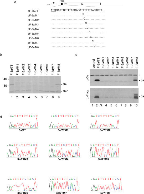
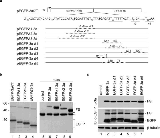
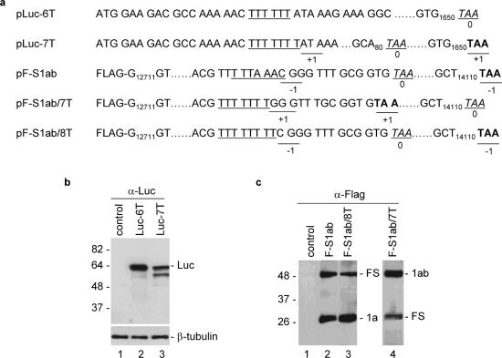
Programmed frameshifting is one of the translational recoding mechanisms that read the genetic code in alternative ways. This process is generally programmed by signals at defined locations in a specific mRNA. In this study, we report the identification of hepta- and octo-uridine stretches as sole signals for programmed +1 and -1 ribosomal frameshifting during translation of severe acute respiratory syndrome coronavirus (SARS-CoV) ORF 3a variants. SARS-CoV ORF 3a encodes a minor structural protein of 274 amino acids. Over the course of cloning and expression of the gene, a mixed population of clones with six, seven, eight and nine T stretches located 14 nt downstream of the initiation codon was found. In vitro and in vivo expression of clones with six, seven and eight Ts, respectively, showed the detection of the full-length 3a protein. Mutagenesis studies led to the identification of the hepta- and octo-uridine stretches as slippery sequences for efficient frameshifting. Interestingly, no stimulatory elements were found in the sequences upstream or downstream of the slippage site. When the hepta- and octo-uridine stretches were used to replace the original slippery sequence of the SARS-CoV ORF 1a and 1b, efficient frameshift events were observed. Furthermore, the efficiencies of frameshifting mediated by the hepta- and octo-uridine stretches were not affected by mutations introduced into a downstream stem-loop structure that totally abolish the frameshift event mediated by the original slippery sequence of ORF 1a and 1b. Taken together, this study identifies the hepta- and octo-uridine stretches that function as sole elements for efficient +1 and -1 ribosomal frameshift events.


Figures

Figure 1
Figure 1
Expression of SARS-CoV ORF 3a variants. (a) Diagram of SARS-CoV 3a constructs with six, seven and eight T stretches under the control of T7 promoter. The positions of six, seven and eight T stretches, the C-terminal His tag in constructs pET3a-His/6T, pET3a-His/7T and pET3a-His/8T, and the N-terminal Flag tag in constructs pF-3a/6T, pF-3a/7T and pF-3a/8T, are indicated. (b) Expression of pSARS-3a/6T (lane 1), pSARS-3a/7T (lane 2) and pSARS-3a/8T (lane 3) in vitro in RRL. Polypeptides were labeled with [35S]methionine, separated on SDS–12% polyacrylamide gel and detected by autoradiography. Bands corresponding to the full-length 3a and a minor species 3a* representing an internal initiation product are indicated. Numbers on the left indicate molecular masses in kilodaltons. (c) Expression of pET3a-His/6T (lane 1), pET3a-His/7T (lane 2) and pET3a-His/8T (lane 3) in bacterial cells. Plasmid DNA was transformed into E.coli strain BL-21, and the protein expression was induced by adding 1 mM IPTG. After induction for 2 h, total cell lysates were prepared, resolved on SDS–12% polyacrylamide, and analyzed by western blot with anti-His antibody. Numbers on the left indicate molecular masses in kilodaltons. (d) Expression of pF-3a/6T (lane 2), pF-3a/7T (lane 3) and pF-3a/8T (lane 4) in Cos-7 cells. Cells were infected with the recombinant vaccinia/T7 virus, and transfected with an empty control plasmid (lane 1) and the three Flag-tagged 3a constructs, respectively. At 18 h posttransfection, cells were harvested and lysates prepared. Polypeptides were separated on SDS–12% polyacrylamide and analyzed by western blot with anti-Flag antibody. Numbers on the left indicate molecular masses in kilodaltons.
Figure 2
Figure 2
Mutational analysis of the slippery sequence in pF-3a/7T. (a) Diagram of pF-3a/7T and the eight mutants (pF-3a/M1-M8). The initiator ATG codon for 3a is underlined. Also shown are the point mutations introduced into the seven T stretch in each mutant constructs. (b) Expression of pF-3a/7T (lane 1) and mutant constructs (lanes 2–9) in vitro in reticulocyte lysates. Polypeptides were labeled with [35S]methionine, separated on SDS–12% polyacrylamide gel and detected by autoradiography. Bands corresponding to the full-length 3a and a minor species 3a* representing an internal initiation product are indicated. Numbers on the left indicate molecular masses in kilodaltons. (c) Expression of pF-3a/7T and mutant constructs in Cos-7 cells. Cells were infected with the recombinant vaccinia/T7 virus, and transfected with an empty control plasmid (lane 1), pF-3a/7T (lane 2) and the eight mutant constructs (lanes 3–10), respectively. At 18 h posttransfection, cells were harvested and lysates prepared. Polypeptides were separated on SDS–12% polyacrylamide and analyzed by western blot with either anti-3a polyclonal (upper panel) or anti-Flag monoclonal antibodies (lower panel). A background band detected by this antiserum is indicated by asterisk. (d) No deletion of nucleotides or revision of mutations in the seven T regions during transcription of wild type and mutant constructs. Cos-7 cells transfected with pF-3a/7T and the eight mutant constructs, respectively, were lysed by Trizol reagent at 24 h post transfection. Total RNA was extracted from cells expressing and 1 µg of RNA was used for RT–PCR. The RT–PCR products were sequenced by automated nucleotide sequencing. The seven T regions of the RT–PCR products are shown.
Figure 3
Figure 3
Deletion analysis of sequences upstream and downstream of the slippage site in pEGFP-3a/7T. (a) Diagram showing the structures of pEGFP-3a/7T and eight derivative constructs with deletion at different regions. Sequence covering the fusion region between EGFP and 3a is shown. The initiator AUG for the 3a ORF is indicated in bold, the position of the nucleotide A is designated +1 and the nucleotide positions upstream and downstream of the AUG codon are indicated by minus and plus numbers, respectively. The seven T stretch is underlined, the termination codon TGA for 0 frame is italic and underlined, and the termination codon TAA for +1 frame is underlined and in bold. Also shown are the nucleotides deleted in each construct. (b) Expression of pEGFP-3a/7T (lanes 1 and 6), pEGFPΔ1-3a (lanes 2 and 7), pEGFPΔ2-3a (lanes 3 and 8) and pEGFPΔ3-3a (lanes 4 and 9) in in vitro reticulocyte lysates (lanes 1–4) and in Cos-7 cells (lanes 5–9). The in vitro expressed polypeptides were labeled with [35S]methionine, separated on SDS–12% polyacrylamide gel and detected by autoradiography. Polypeptides expressed in Cos-7 cells were separated on SDS–12% polyacrylamide and analyzed by western blot with anti-3a polyclonal antibodies. Bands corresponding to EGFP, the frameshifting products (FS) and a background band (asterisk) are indicated. Numbers on the left indicate molecular masses in kilodaltons. (c) Expression of pEGFP-3a/7T (lane 2), pEGFP-3aΔ1 (lane 3), pEGFP-3aΔ2 (lane 4) pEGFP-3aΔ3 (lane 5), pEGFP-3aΔ4 (lane 6) and pEGFP-3aΔ5 (lane 7) in Cos-7 cells. Polypeptides expressed were separated on SDS–12% polyacrylamide and analyzed by western blot with either anti-3a, anti-EGFP, or anti-β-tubulin antibodies. Bands corresponding to EGFP, the frameshifting products and -β-tubulin are indicated.
Figure 4
Figure 4
Analysis of frameshifting efficiencies mediated by hepta- and octo-uridine stretches in heterogeneous ORFs. (a) Diagram showing the structures of constructs pLuc-6T, pLuc-7T, pF-S1ab, pF-S1ab/7T and pF-S1ab/8T. The slippery sequences (the six T stretch in the case of pLuc-6T) is underlined, the TAA termination codon for 0 frame is italic and underlined, and the TAA termination codon for +1 or −1 frame is underlined and in bold. (b) Expression of pLuc-6T and pLuc-7T in Cos-7 cells. Cells were infected with the recombinant vaccinia/T7 virus, and transfected with an empty control plasmid (lane 1), pLuc-6T (lane 2) and pLuc-7T (lane 3). At 18 h posttransfection, cells were harvested and lysates prepared. Polypeptides were separated on SDS–12% polyacrylamide and analyzed by western blot with anti-luciferase antibodies. Bands corresponding to the full-length luciferase and -β-tubulin are indicated. Numbers on the left indicate molecular masses in kilodaltons. (c) Expression of pF-S1ab, pF-S1ab/7T and pF-S1ab/8T in Cos-7 cells. Cells were infected with the recombinant vaccinia/T7 virus, and transfected with an empty control plasmid (lane 1), pF-S1ab (lane 2), pF-S1ab/7T (lane 3) and pF-S1ab/8T (lane 4). At 18 h posttransfection, cells were harvested and lysates prepared. Polypeptides were separated on SDS–12% polyacrylamide and analyzed by western blot with anti-Flag antibodies. Bands corresponding to 1a (or 1ab) and the frameshifting products are indicated. Numbers on the left indicate molecular masses in kilodaltons.
Figure 5
Figure 5
Mutational analysis of a downstream stimulator on frameshifting efficiencies mediated by the hepta- and octo-uridine stretches. (a) Diagram showing the slippery sequence and the downstream stem–loop structures of SARS-CoV 1a/1b region. The slippery sequence is underlined and the two stems are indicated. Also indicated are the mutations introduced into stem 2 (in bold). (b) Expression of pF-S1ab (lane 1), pF-S1abM (lane 2), pF-S1ab/7T (lane 6), pF-S1ab/7TM (lane 7), pF-S1ab/8T (lane 3) and pF-S1ab/8T (lane 4) in Cos-7 cells. Cells were infected with the recombinant vaccinia/T7 virus, and transfected with an empty control plasmid (lane 5) and the six constructs, respectively. At 18 h posttransfection, cells were harvested and lysates prepared. Polypeptides were separated on SDS–12% polyacrylamide and analyzed by western blot with anti-Flag antibodies. Bands corresponding to 1a (or 1ab), the frameshifting products (FS) and β-tubulin are indicated. Numbers on the left indicate molecular masses in kilodaltons.
Figure 6
Figure 6
Differential effects of a downstream stimulator on frameshifting efficiencies mediated by wild type and mutant hepta-uridine stretch. (a) Diagram showing the structure of constructs pF-S1ab/7T, pF-S1ab/7TM1C, pF-S1ab/7TM1A, pF-S1ab/7TM7C and pF-S1ab/7TM14C. The seven T stretch is underlined. Also shown are the TAA termination codon for the +1 and 0 frames, and the mutations introduced into the seven T region. (b) Expression of pF-S1ab/7T (lane 2), pF-S1ab/7TM1A (lane 3), pF-S1ab/7TM1C (lane 4), pF-S1ab/7TM7C (lane 5) and pF-S1ab/7TM14C (lane 6) in Cos-7 cells. Cells were infected with the recombinant vaccinia/T7 virus and infected either with an empty control plasmid (lane 1) or each of the five constructs (lanes 2–6). At 18 h posttransfection, cells were harvested and lysates prepared. Polypeptides were separated on SDS–12% polyacrylamide gel and analyzed by western blot with anti-Flag antibodies. Bands corresponding to 1ab, the frameshifting products (FS) and β-tubulin are indicated. Numbers on the left indicate molecular masses in kilodaltons.

Similar articles

Cited by

References

    1. Belcourt M.F., Farabaugh P.J. Ribosomal frameshifting in the yeast retrotransposon Ty: tRNAs induce slippage on a 7 nucleotide minimal site. Cell. 1990;62:339–352. - PMC - PubMed
    1. Farabaugh P.J., Zhao H., Vimaladithan A. A novel programmed frameshift expresses the POL3 gene of retrotransposon Ty3 of yeast: frameshifting without tRNA slippage. Cell. 1993;74:93–103. - PMC - PubMed
    1. Blinkowa A.L., Walker Programmed ribosomal frameshifting generates the Escherichia coli DNA polymerase III γ subunit from within the τ subunit reading frame. Nucleic Acids Res. 1990;18:1725–1729. - PMC - PubMed
    1. Craigen W.J., Caskey C.T. Expression of peptide chain release factor 2 requires high-efficiency frameshift. Nature. 1986;322:273–275. - PubMed
    1. Gurvich O., Baranov P.V., Zhou J., Hammer A.W., Gesteland R., Atkins J.F. Sequences that direct significant levels of frameshifting are frequent in coding regions of Escherichia coli. EMBO J. 2003;22:5941–5950. - PMC - PubMed

Publication types