

Three structural roles for water in bone observed by solid-state NMR
- PMID: 16500963
- PMCID: PMC1440753
- DOI: 10.1529/biophysj.105.070243
Three structural roles for water in bone observed by solid-state NMR
Abstract
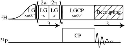
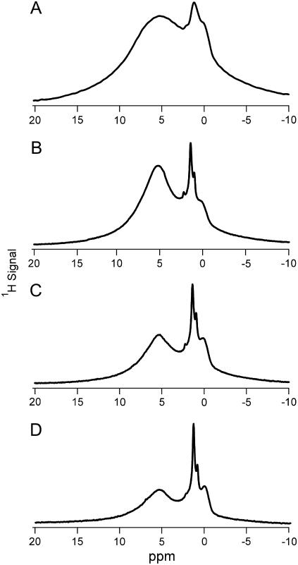
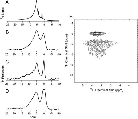
Hydrogen-bearing species in the bone mineral environment were investigated using solid-state NMR spectroscopy of powdered bone, deproteinated bone, and B-type carbonated apatite. Using magic-angle spinning and cross-polarization techniques three types of structurally-bound water were observed in these materials. Two of these water types occupy vacancies within the apatitic mineral crystal in synthetic carbonated apatite and deproteinated bone and serve to stabilize these defect-containing crystals. The third water was observed at the mineral surface in unmodified bone but not in deproteinated bone, suggesting a role for this water in mediating mineral-organic matrix interactions. Direct evidence of monohydrogen phosphate in a (1)H NMR spectrum of unmodified bone is presented for the first time. We obtained clear evidence for the presence of hydroxide ion in deproteinated bone by (1)H MAS NMR. A (1)H-(31)P heteronuclear correlation experiment provided unambiguous evidence for hydroxide ion in unmodified bone as well. Hydroxide ion in both unmodified and deproteinated bone mineral was found to participate in hydrogen bonding with neighboring water molecules and ions. In unmodified bone mineral hydroxide ion was found, through a (1)H-(31)P heteronuclear correlation experiment, to be confined to a small portion of the mineral crystal, probably the internal portion.
Figures

References
-
- Elliott, J. C. 2002. Calcium phosphate biominerals. In Reviews in Mineralogy & Geochemistry, Vol. 48. M. L. Kohn, J. Rakovan, and T. M. Hughes, editors. Minerology Society of America, Washington, DC. 427–453.
-
- Sudarsanan, K., and R. A. Young. 1969. Significant precision in crystal structural details: Holly Springs hydroxyapatite. Acta Crystallogr. B. 25:1534–1543.
-
- Ivanova, T. I., O. V. Frank-Kamenetskaya, A. B. Kol'tsov, and V. L. Ugolkov. 2001. Crystal structure of calcium-deficient carbonated hydroxyapatite. Thermal decomposition. J. Solid State Chem. 160:340–349.
-
- Loong, C.-K., C. Rey, L. T. Kuhn, C. Combes, Y. Wu, S.-H. Chen, and M. J. Glimcher. 2000. Evidence of hydroxyl-ion deficiency in bone apatites: an inelastic neutron-scattering study. Bone. 26:599–602. - PubMed
-
- Rey, C., J. L. Miquel, L. Facchini, A. P. Legrand, and M. J. Glimcher. 1995. Hydroxyl groups in bone mineral. Bone. 16:583–586. - PubMed
Publication types
MeSH terms
Grants and funding
LinkOut - more resources
Full Text Sources
