. 2006 Apr;140(4):1507-21.
doi: 10.1104/pp.105.070672. Epub 2006 Feb 24.

Calmodulin interacts with and regulates the RNA-binding activity of an Arabidopsis polyadenylation factor subunit

Affiliations

Calmodulin interacts with and regulates the RNA-binding activity of an Arabidopsis polyadenylation factor subunit

Kimberly J Delaney et al. Plant Physiol. 2006 Apr.

Abstract

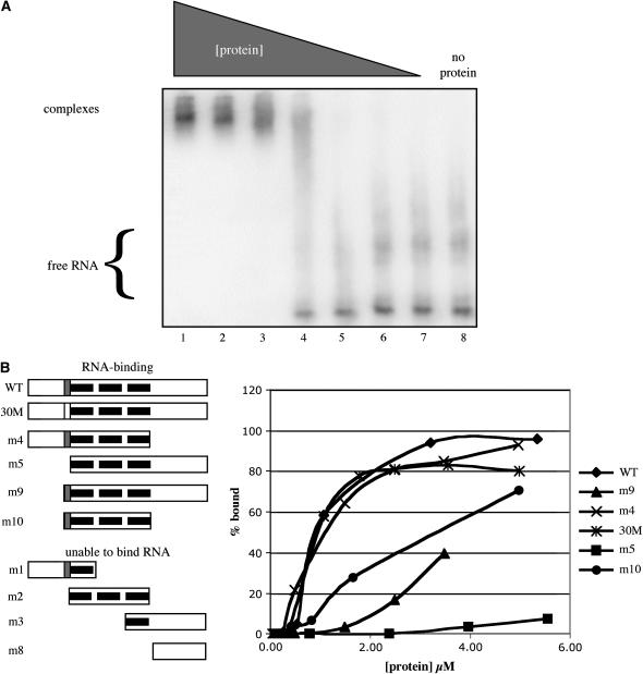
The Arabidopsis (Arabidopsis thaliana) gene that encodes the probable ortholog of the 30-kD subunit of the mammalian cleavage and polyadenylation specificity factor (CPSF) is a complex one, encoding small (approximately 28 kD) and large (approximately 68 kD) polypeptides. The small polypeptide (AtCPSF30) corresponds to CPSF30 and is the focus of this study. Recombinant AtCPSF30 was purified from Escherichia coli and found to possess RNA-binding activity. Mutational analysis indicated that an evolutionarily conserved central core of AtCPSF30 is involved in RNA binding, but that RNA binding also requires a short sequence adjacent to the N terminus of the central core. AtCPSF30 was found to bind calmodulin, and calmodulin inhibited the RNA-binding activity of the protein in a calcium-dependent manner. Mutational analysis showed that a small part of the protein, again adjacent to the N terminus of the conserved core, is responsible for calmodulin binding; point mutations in this region abolished both binding to and inhibition of RNA binding by calmodulin. Interestingly, AtCPSF30 was capable of self-interactions. This property also mapped to the central conserved core of the protein. However, calmodulin had no discernible effect on the self-association. These results show that the central portion of AtCPSF30 is involved in a number of important functions, and they raise interesting possibilities for both the interplay between splicing and polyadenylation and the regulation of these processes by stimuli that act through calmodulin.


Figures

Figure 1.
Figure 1.
Properties of the Arabidopsis gene (At1g30460) that encodes AtCPSF30. A, Sequence alignments of AtCPSF30 with other eukaryotic CPSF30 proteins. Amino acid identity is denoted with white uppercase letters on a black background, similarity with black uppercase letters on a gray background, and unrelated sequences with lowercase lettering. The “N,” “Z,” and “C” domains mentioned in the text are denoted above the Arabidopsis sequence, and are delimited with a “|” symbol above the residues that demarcate the three domains. The domain identified by calmodulin-binding domain prediction programs (http://calcium.uhnres.utoronto.ca/ctdb/ctdb/sequence.html) is underlined, and the four amino acids that were changed to Ala in the 30M mutant are indicated with a lowercase “a” beneath the respective position. GenBank accessions for the eukaryotic sequences are as follows: mammalian (Bos taurus), AAC48759; Drosophila (Drosophila melanogaster), AAF51453; and yeast (Saccharomyces cerevisiae), NP_015432. B, Diagram of the structure of the gene, showing the intron/exon organization (introns are thin lines, exons thick ones), and the two transcripts that encode AtCPSF30 and AtCPSF30-YT521-B, respectively. GenBank accessions for plant-derived expressed sequence tags that correspond to the two transcripts are listed beneath each one. The position of the T-DNA insertion in the oxt6 mutant is indicated above the respective exon; this site is 147 bp 3′ of the translation initiation codon for this gene.
Figure 2.
Figure 2.
At1g30460-encoded proteins are in nuclear complexes. A, Expression of the gene encoding AtCPSF30. Total RNA from wild-type and oxt6 mutant plants (see Fig. 1 for the location of the T-DNA insert in this mutant) were analyzed by RNA blotting using a probe derived from the first exon (top) or by RT-PCR, using primers specific for the smaller of the two transcripts (middle). At1g30460-derived transcripts are denoted as “1” and “2,” respectively. The RNA blot was also probed with tubulin sequences (bottom). B, At1g30460-encoded polypeptides can be detected in Arabidopsis nuclear extracts. Immunoblot filters containing separated Arabidopsis nuclear extracts were probed with affinity-purified anti-AtCPSF30 antibody. Protein size markers are on the left lane, and the arrowheads indicate expected bands that correspond to the products of the small and large At1g30460-encoded mRNAs. Nuclear extracts from the oxt6 mutant (lane 1) and the wild type (lane 2) were used to discriminate two expected proteins encoded by the two mRNAs of At1g30460. The smaller band corresponds to AtCPSF30. C, Coimmunoprecipitation. Equal amounts of Arabidopsis nuclear protein extracts were precipitated by the affinity-purified anti-AtCPSF100 antibody (lane marked “Antibody”) and preimmune antiserum (lane marked “Preimmune”), respectively. AtCPSF30 (pointed by arrowhead) was detected from the pellet immunoprecipitated by anti-AtCPSF100 antibody.
Figure 3.
Figure 3.
Amino acid sequence comparisons of CCCH proteins. A tree diagram is shown that summarizes the amino acid sequences similarities of plant CPSF30-like proteins with other eukaryotic CPSF30s and with the set of Arabidopsis CCCH zinc-finger proteins. The plant and other eukaryotic CPSF30 sequences are highlighted with shaded boxes as indicated. Putative Arabidopsis CCCH motif-containing proteins were identified by BLAST searches; those analyzed are At1g30460 (= AtCPSF30), At1g21570, At5g56930, At2g47680, At3g47120, At1g66810, At3g08505, At3g08505, At5g49200, At4g25440, At1g04990, At1g04990, At5g63260, At2g19810, At1g32360, At2g32930, At5g16540, At5g16540, At5g16540, At2g25900, At5g56900, At2g35430, At5g56900, At5g51980, At3g02830, At5g40880, At1g48195, At1g67460, At1g32975, At2g02160, At2g47850, At1g15100, At4g29190, At3g55980, At3g19360, At2g05160, At1g68200, At5g12850, At2g40140, At3g06410, At2g36040, At3g12130, At3g44785, At4g26850, At3g48440, At5g18550, At1g75340, At1g27650, and At3g05730. GenBank accessions for the other eukaryotic sequences in this analysis were as follows: human, EAL23878; B. taurus, AAC48759; Drosophila (D. melanogaster), AAF51453; Danio (Danio rerio), AAH45289; S. cerevisiae, NP_015432; Schizosaccharomyces pombe, CAB61457; poplar (Populus spp.), CV243319; and Nicotiana, CK286112.
Figure 4.
Figure 4.
Sequence alignments of AtCPSF30 with other plant CPSF30-like proteins. Amino acid identity is denoted with white uppercase letters on a black background, similarity with black uppercase letters on a gray background, and unrelated sequences with lowercase lettering. The central zinc-finger core that is shared with eukaryotic CPSF30 proteins is highlighted with a dashed underline, and the N- and C-terminal limits of this core are denoted with a “|” beneath the sequence. The thin vertical bar beneath the first line of the Arabidopsis sequence denotes the endpoint of the m6 mutation (see Fig. 9). The thick vertical bar beneath the last line of the Arabidopsis sequence shows the location of the splice site that is used in the biogenesis of the larger At1g30460-derived mRNA; amino acid sequences to the C-terminal side of this are absent from the AtCPSF30-YT521-B polypeptide. Accessions are given in the legend for Figure 2.
Figure 5.
Figure 5.
Demarcation of the RNA-binding domain of AtCPSF30. A, Binding of RNA by wild-type AtCPSF30 as a function of protein concentration. A total of 3.5 pmol of uniformly labeled RNA containing the polyadenylation signal of the pea rbcS-E9 gene was incubated with varying quantities of MBP-AtCPSF4 and RNA binding assessed by electrophoresis on native gels and autoradiography. The positions of the RNA-protein complex (complexes) and free RNA are indicated on the left. No protein was added for the sample in lane 8. Lanes 1 to 7 contained 10.5, 7.5, 4.5, 1.5, 0.75, 0.375, and 0.188 μg of purified protein, respectively. B, RNA binding by mutant forms of AtCPSF30. On the left is a depiction of the different variants of AtCPSF30 that were produced as MBP fusion proteins. The central zinc-finger domain is represented with three black lines within the respective rectangular boxes, and the small domain that is responsible for calmodulin binding shown as a gray box within the larger representation. The clear box in the 30M representation indicates that the calmodulin-binding motif has been changed by mutation to be nonfunctional. Fusion proteins that bound RNA are listed under “RNA-binding,” and the plots of binding as a function of protein concentration given on the right. Fusion proteins for which no detectable RNA binding was observed are listed under “unable to bind RNA”; the plots for these proteins are not given, as they would coincide with the x axis.
Figure 6.
Figure 6.
Identification of the calmodulin-binding domain of AtCPSF30. Proteins that were analyzed for calmodulin binding are depicted on the left; the explanation of the depiction is as in the legend for Figure 5. The results of SDS-PAGE and staining (top; labeled “stained gel”) and far-western blotting using biotinylated calmodulin (bottom; labeled “calmodulin-binding”) are shown. 30M-4, 30M-5, and 30M-6 are preparations from three independent mutants in which the putative calmodulin-binding domain was altered as shown on the left (altered amino acids are underlined). The 30M-4 preparation was used in the other studies in this report.
Figure 7.
Figure 7.
Effects of calmodulin on RNA binding by AtCPSF30 and the 30M mutant. A total of 1.25 pmol of uniformly labeled RNA containing the polyadenylation signal of the pea rbcS-E9 gene was incubated with 40 pmol of MBP-AtCPSF30 (WT) or the purified 30M mutant protein (MUT), and with various combinations of calmodulin (60 pmol; CAM), calcium chloride (10 μm), and EGTA (1 mm), and RNA binding assessed by electrophoresis on native gels and autoradiography. The ratios of binding activity observed in the presence or absence of calmodulin (activity + CAM/activity − CAM) were plotted for each of four sets of conditions: wild-type protein + calcium chloride (WT + Ca), mutant protein + calcium chloride (MUT + Ca), wild-type protein + EGTA (WT + EGTA), and the mutant protein + EGTA (MUT + EGTA). Purified calmodulin does not bind RNA in this assay and has no effect on the added RNA (data not shown).
Figure 8.
Figure 8.
AtCPSF30 interacts with itself. A, Depiction of the AtCPSF30 variants that were produced as labeled proteins. The symbols used are described in the legend for Figure 5. B, Binding of labeled AtCPSF30 and deletion derivatives to MBP-AtCPSF30 and MBP. Experiments were performed as described in “Materials and Methods.” The identity of the labeled protein is indicated at the top. Gels showing results with MBP-AtCPSF30 (top) or MBP (middle) as baits are shown. Ten percent of the quantity of each translation mixture used in the copurification assays is shown in the bottom section [“onto” (10%)]. C, Calmodulin has no effect on the self-interaction. Copurification assays with labeled AtCPSF30 and various combinations of calmodulin, CaCl2, and EGTA were performed. Lane 1, 10% of the labeled input protein. Lanes 2 to 6, All had the same labeled input (AtCPSF30). Lane 2, No addition. Lanes 3 and 5, 2.5 mm Ca Cl2. Lanes 4 and 6, 2.5 mm EGTA. Lanes 5 and 6, + calmodulin.
Figure 9.
Figure 9.
A functional map of AtCPSF30. The complete AtCPSF30 coding region is represented as the long, lightly shaded rectangle. The three zinc-finger motifs are indicated with thick black lines lying within the AtCPSF30 coding region. Above this box are shown the relative locations of the domains responsible for self-association (CPSF30) and RNA binding (RNA); shading near the ends of these boxes indicates uncertainties in the possible extents of these two domains. The location of the calmodulin-binding site is shown beneath the light gray box (C).

Similar articles

Cited by

References

    1. Addepalli B, Meeks LR, Forbes KP, Hunt AG (2004) Novel alternative splicing of mRNAs encoding poly(A) polymerases in Arabidopsis. Biochim Biophys Acta 1679: 117–128 - PubMed
    1. Altschul SF, Madden TL, Schaffer AA, Zhang J, Zhang Z, Miller W, Lipman DJ (1997) Gapped BLAST and PSI-BLAST: a new generation of protein database search programs. Nucleic Acids Res 25: 3389–3402 - PMC - PubMed
    1. Amasino RM (2003) Flowering time: a pathway that begins at the 3′ end. Curr Biol 13: 670–672 - PubMed
    1. Bai C, Tolias PP (1998) Drosophila clipper/CPSF 30K is a post-transcriptionally regulated nuclear protein that binds RNA containing GC clusters. Nucleic Acids Res 26: 1597–1604 - PMC - PubMed
    1. Barabino SM, Hubner W, Jenny A, Minvielle-Sebastia L, Keller W (1997) The 30-kD subunit of mammalian cleavage and polyadenylation specificity factor and its yeast homolog are RNA-binding zinc finger proteins. Genes Dev 11: 1703–1716 - PubMed

Publication types

MeSH terms

Associated data