. 2006 Mar 7;103(10):3627-32.
doi: 10.1073/pnas.0509929103. Epub 2006 Feb 24.

Projection structure of the human copper transporter CTR1 at 6-A resolution reveals a compact trimer with a novel channel-like architecture

Affiliations

Projection structure of the human copper transporter CTR1 at 6-A resolution reveals a compact trimer with a novel channel-like architecture

Stephen G Aller et al. Proc Natl Acad Sci U S A. .

Abstract

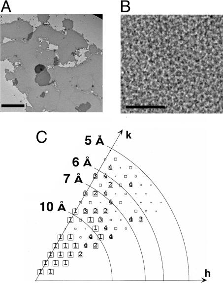
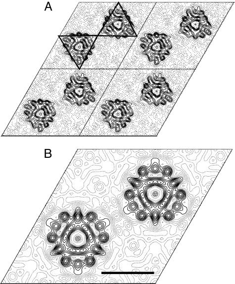
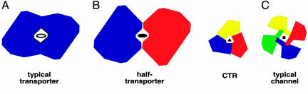
Human CTR1 is a high-affinity copper transporter that also mediates the uptake of the anticancer drug cisplatin by largely unknown transport mechanisms. Here we report the 6-A projection structure obtained for human CTR1 by using electron crystallography of 2D protein crystals in a native phospholipid bilayer. The projection of CTR1 reveals a symmetrical trimer that is <40 A wide. Notably, the center threefold axis of each trimer forms a region of very low electron density likely to be involved in copper translocation. The formation of a putative pore for metal ions at the interface of three identical subunits deviates from the structural design of typical primary and secondary active transporters and reveals that copper uptake transporters have a novel architecture that is structurally more closely related to channel proteins.


Conflict of interest statement

Conflict of interest statement: No conflicts declared.

Figures

Fig. 1.
Fig. 1.
N-terminal HA-tagged hCTR1N15Q construct is functional in growth complementation under copper starvation. S. cerevisiae yeast deficient in high-affinity copper uptake (Δctr1,3) were transformed with the HA-tagged hCTR1 construct used in this study (labeled as HAhCTR1N15Q) or vector only (vector). (Upper) Copper starvation was achieved with plates that contained the copper chelator bathocuproinedisulfonic acid (BCS) and no additional copper. (Lower) Shown is control growth on plates containing excess copper sulfate (50 μM) without any chelator.
Fig. 2.
Fig. 2.
Gel filtration of purified HAhCTR1N15Q reveals two disulfide-dependent oligomerization states. (A) HAhCTR1N15Q in the absence of reducing agents. (B) HAhCTR1N15Q chromatographed after the column was preequilibrated with buffer containing the disulfide-specific reducing agent TCEP (1 mM). In the absence of TCEP, a HAhCTR1N15Q dimer-of-trimer species eluted in fraction 10 and the trimer eluted in fraction 13. (C) Coomassie staining of 10 μl each of gel filtration fractions 7–15 and the same fractions diluted 1:1,000 subjected to anti-HA Western blot. Fractions 10 and 13 are specifically indicated. The smear observed below the band of the dimer most likely reflects anomalous migration caused by flexibility of the dimeric species.
Fig. 3.
Fig. 3.
Crosslinking of purified HAhCTR1N15Q. Gel filtration fraction 10 was incubated in the indicated concentrations of primary-amine reactive crosslinkers of varying spacer lengths (1,5-difluoro-2,4-dinitrobenzene, DFDNB, 3 Å; dithiobis-[succinimidylpropionate], DSP, 12 Å; ethylene glycolbis[succinimidylsuccinate], EGS, 16 Å) for 30 min before quenching the reaction and subjection to nonreducing SDS/PAGE and Coomassie staining. The apparent increase in weight likely reflects the differences in molecular mass of the crosslinking agents themselves, but could also be related to slight differences in the compactness of the crosslinked oligomer.
Fig. 4.
Fig. 4.
2D crystals of purified HAhCTR1N15Q. (A) Low-magnification view of flat sheets of HAhCTR1N15Q crystals stained with 2% phosphotungstic acid. (Scale bar corresponds to 1.5 μm.) (B) High-magnification view (×37,000) of the hexagonal lattice. (Scale bar corresponds to 500 Å.) (C) Phase error of unique reflections to 5-Å resolution. The size of the boxes in the plot correspond to the phase error associated with each measurement after averaging data from five images and rounding to 0° or 180° as dictated by the centrosymmetric constraint (1, <8°; 2, <14°; 3, <20°; 4, <30°; 5, <40°; 6, <50°; 7, <80°; 8,<90°, where 90° is random). Boxes are shown as decreasing in size depending on the phase error, and sizes 1–4 are individually labeled.
Fig. 5.
Fig. 5.
Projection views of hCTR1. (A) Projection density map of HAhCTR1N15Q at 6-Å resolution with no symmetry enforced (p1). Four unit cells (a = b = 87.9 Å) are shown. Triangles are drawn around two of the superimposed dimers of trimers in a single unit cell to emphasize the triangular shape of the complex. (B) A single unit cell is displayed with p622 symmetry imposed. The double-layered crystals have four hCTR1 trimers in each unit cell. Strong circular density features are indicative of α-helical secondary structure but cannot be unambiguously attributed to the three transmembrane domains. A region of very low electron density at the center axis of each hCTR1 trimer marks the presence of a putative copper permeable pore within the complex. (Scale bar corresponds to a distance of 30 Å.)
Fig. 6.
Fig. 6.
3D reconstruction of HAhCTR1N15Q. A stereo side view of half a unit cell (two stacked trimers) is shown with p6 symmetry imposed. Because p6 symmetry cannot impose a second layer during data processing, the two layers are truly crystallographic. Thus, enforcing p622 symmetry for the calculated projection structure (Fig. 5) is justified. A distance of 30 Å for the hydrophobic acyl chains of each phospholipid bilayer is marked to scale as Mh. The total distance of each phospholipid bilayer including lipid head groups (≈45 Å) is marked Mt. The placement of these boundaries is an approximation. The plot is contoured at 1.2 σ.
Fig. 7.
Fig. 7.
hCTR1 is organized like a transporter-channel hybrid. (A) In the majority of known transporter structures, the permeation pathway is contained within a single monomer that displays pseudo twofold symmetry. This architecture is seen for the structures of the multidrug efflux transporter AcrB, the ammonia transporter AmtB, the E. coli H+/Cl exchange transporter, the glycerol-3-phosphate transporter GlpT, the lactose permease LacY, the Na+/Cl-dependent leucine transporter LeuT, and the oxalate transporter OxlT. In three cases, even though the pseudo twofold axis of symmetry is not evident, the monomeric unit still forms an independent pore (glutamate transporter Glt, NhaA, and the calcium pump of sarcoplasmic reticulum). (B) Half-transporters of the ATP-binding cassette family, such as MsbA and BtuCD, have 6–10 transmembrane domains. Although these helical bundles would be large enough to accommodate a pore within the monomer, these transporters homodimerize, forming a pore for substrates at the interface of monomers. In contrast, hCTR1 monomers are too small to form a pore, thus making oligomerization mandatory for function. (C) Our data support that the pore for copper ions is formed at the interface of three identical subunits that oligomerize in a manner much like traditional channel proteins.

References

    1. Fu X., Rinaldo P., Hahn S. H., Kodama H., Packman S. J. Inherited Metab. Dis. 2003;26:55–66. - PubMed
    1. Lee J., Prohaska J. R., Thiele D. J. Proc. Natl. Acad. Sci. USA. 2001;98:6842–6847. - PMC - PubMed
    1. Kuo Y. M., Zhou B., Cosco D., Gitschier J. Proc. Natl. Acad. Sci. USA. 2001;98:6836–6841. - PMC - PubMed
    1. Ishida S., Lee J., Thiele D. J., Herskowitz I. Proc. Natl. Acad. Sci. USA. 2002;99:14298–14302. - PMC - PubMed
    1. Lin X., Okuda T., Holzer A., Howell S. B. Mol. Pharmacol. 2002;62:1154–1159. - PubMed

Publication types

MeSH terms