. 2006 Mar 7;103(10):3758-63.
doi: 10.1073/pnas.0508917103. Epub 2006 Feb 24.

Body-wide gene therapy of Duchenne muscular dystrophy in the mdx mouse model

Affiliations

Body-wide gene therapy of Duchenne muscular dystrophy in the mdx mouse model

Michela Alessandra Denti et al. Proc Natl Acad Sci U S A. .

Abstract

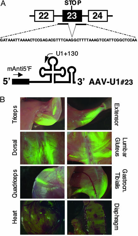
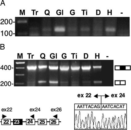
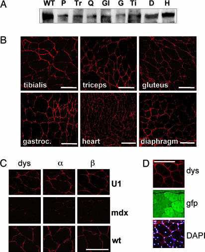
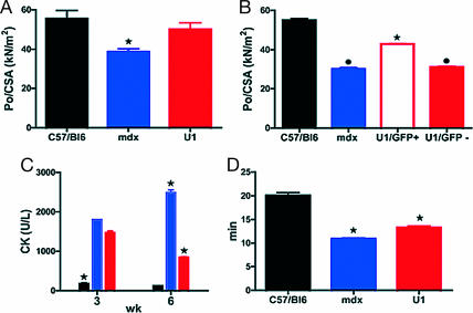
Duchenne muscular dystrophy is an X-linked muscle disease characterized by mutations in the dystrophin gene. Many of these can be corrected at the posttranscriptional level by skipping the mutated exon. We have obtained persistent exon skipping in mdx mice by tail vein injection with an adeno-associated viral (AAV) vector expressing antisense sequences as part of the stable cellular U1 small nuclear RNA. Systemic delivery of the AAV construct resulted in effective body-wide colonization, significant recovery of the functional properties in vivo, and lower creatine kinase serum levels, suggesting an overall decrease in muscle wasting. The transduced muscles rescued dystrophin expression and displayed a significant recovery of function toward the normal values at single muscle fiber level. This approach provides solid bases for a systemic use of AAV-mediated antisense-U1 small nuclear RNA expression for the therapeutic treatment of Duchenne muscular dystrophy.


Conflict of interest statement

Conflict of interest statement: No conflicts declared.

Figures

Fig. 1.
Fig. 1.
AAV2/1 efficiently delivers antisense constructs to muscles. (A) Schematic representation of the mdx dystrophin mutation and of the AAV-U1#23 antisense construct. Antisense sequences complementary to the 5′ and 3′ splice sites of exon 23 (black bars) were cloned consecutively in the 5′ portion of U1 snRNA (black box). The corresponding antisense sequence is shown underneath the dystrophin pre-mRNA. (B) Muscle transduction after AAV2/1 systemic delivery. Green fluorescence was evident in several muscle districts 12 weeks after tail vein administration of 3–4 × 10¹² genome copies/mouse of AAV-U1#23. Intensively transduced fiber groups flank less or nonfluorescent muscle fibers possibly reflecting heterogeneous perfusion inside the muscle. Heart and diaphragm are also widely transduced although to a lower level than voluntary striated muscle.
Fig. 2.
Fig. 2.
Analysis of antisense construct expression and exon 23 skipping. (A) Expression of chimeric antisense U1 construct in body-wide muscles 12 weeks after tail vein injection of the AAV-U1#23 construct. Nested RT-PCR was performed on 200 ng of total RNA with primers U1 + 130 and mAnti5′F (Fig. 1A). The products were run on a 2% agarose gel. Lanes were as follows: M, 100-bp DNA ladder (Invitrogen); Tr, triceps; Q, quadriceps; Gl, gluteus; G, gastrocnemius; Ti, tibialis; D, diaphragm; H, heart; −, RNA-minus control. (B) Skipping of exon 23. In the schematic representation of a portion of the dystrophin pre-mRNA, the oligos used for the nested RT-PCRs are indicated. Lanes are as in A. The chromatogram indicates that correct fusion between exon 22 and 24 has occurred. A schematic representation of the unskipped and skipped products is shown on the side of the gel.
Fig. 3.
Fig. 3.
Analysis of dystrophin and DAPC rescue. (A) Two hundred micrograms of total proteins from different muscles were analyzed 12 weeks after injection by Western blotting with anti-dystrophin antibodies in parallel with 5 μg of proteins extracted from WT skeletal muscle cells (lane WT). P, pectoralis; Tr, triceps; Q, quadriceps; Gl, gluteus; G, gastrocnemius; Ti, tibialis; D, diaphragm; H, heart. (B) The same muscles were also sliced and analyzed by immunofluorescence with anti-dystrophin antibodies. Pictures were taken with a confocal microscope. (C) Sections of the quadriceps of AAV-U1#23-injected mice were also reacted with anti-α- and -β-sarcoglycan antibodies. Middle and Bottom panels indicate the reactivity of quadriceps sections of an untreated mdx mouse and of a WT mouse, respectively, with the same antibodies as above. Pictures were taken with a confocal microscope. (D) Rescue of dystrophin synthesis (dys) corresponded to the expression of EGFP protein (gfp). (DAPI) Bottom panel shows the merging of dystrophin signals with those of the DAPI staining. (Scale bars: 100 μm.)
Fig. 4.
Fig. 4.
Functional tests on mdx mice injected systemically with AAV-U1#23. Controls (black), untreated mdx (blue), and AAV-U1#23 (red)-treated mdx mice. (A) Specific force of single fibers from the gastrocnemius (n = 120, mice n = 3 for each group) was significantly lower in mdx mice in comparison with controls (P ≤ 0.01) and recovered toward normal values in AAV-U1#23 (+30%, P ≤ 0.01). (B) Po/CSA of fibers (n = 150, mice n = 3 for each group) from the vastus of C57BL/6 and mdx and from small GFP-positive and GFP-negative bundles of AAV-U1#23-treated muscles. GFP-positive fibers from treated mice showed Po/CSA values significantly higher (P ≤ 0.001) than those obtained from GFP-negative fibers of treated mice and fibers of untreated mdx mice (P ≤ 0.001). (C) CKnac (units/liter) serum levels were monitored 3 and 6 weeks after AAV-U1#23 injections in parallel to C57BL/6 and mdx mice. (D) Untreated mdx mice showed a significant reduction (P ≤ 0.001) of the tolerance to treadmill exhaustion test compared with C57BL/6, whereas a partial but significant recovery was observed after AAV-U1#23 treatment (P ≤ 0.01). ∗, significantly different from the other groups; bullet, significantly different from C57BL/6 and AAV-U1#23 GFP-positive. Data are mean values ± SEM. Statistical analysis was assessed by ANOVA followed by the Student–Newman–Keuls test. P ≤ 0.05 was considered significant.

References

    1. Koenig M., Beggs A. H., Moyer M., Scherpf S., Heindrich K., Bettecken T., Meng G., Muller C. R., Lindlof M., Kaariainen H., et al. Am. J. Hum. Genet. 1989;45:498–506. - PMC - PubMed
    1. Sicinski P., Geng Y., Ryder-Cook A. S., Barnard E. A., Darlison M. G., Barnard P. J. Science. 1989;244:1578–1580. - PubMed
    1. DelloRusso C., Scott J. M., Hartigan-O’Connor D., Salvatori G., Barjot C., Robinson A. S., Crawford R. W., Brooks S. V., Chamberlain J. S. Proc. Natl. Acad. Sci. USA. 2002;99:12979–12984. - PMC - PubMed
    1. Jiang Z., Schiedner G., van Rooijen N., Liu C. C., Kochanek S., Clemens P. R. Mol. Ther. 2004;10:688–696. - PubMed
    1. Wang B., Li J., Xiao X. Proc. Natl. Acad. Sci. USA. 2000;97:13714–13719. - PMC - PubMed

Publication types