. 2006 Mar;80(6):2596-608.
doi: 10.1128/JVI.80.6.2596-2608.2006.

Epitope mapping of herpes simplex virus type 2 gH/gL defines distinct antigenic sites, including some associated with biological function

Affiliations

Epitope mapping of herpes simplex virus type 2 gH/gL defines distinct antigenic sites, including some associated with biological function

Tina M Cairns et al. J Virol. 2006 Mar.

Abstract

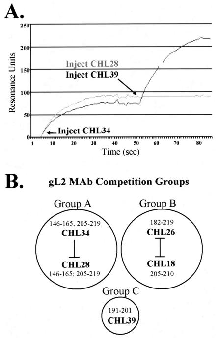
The gH/gL complex plays an essential role in virus entry and cell-cell spread of herpes simplex virus (HSV). Very few immunologic reagents were previously available to either identify important functional regions or gain information about structural features of this complex. Therefore, we generated and characterized a panel of 31 monoclonal antibodies (MAbs) against HSV type 2 (HSV-2) gH/gL. Fourteen MAbs bound to a conformation-dependent epitope of the gH2/gL2 complex, and all blocked virus spread. The other 17 MAbs recognized linear epitopes of gH (12) or gL (5). Interestingly, two of the gL MAbs and six of the gH MAbs were type common. Overlapping synthetic peptides were used to map MAbs against linear epitopes. These data, along with results of competition analyses and functional assays, assigned the MAbs to groups representing eight distinct antigenic sites on gH (I to VIII) and three sites on gL (A, B, and C). Of most importance, the MAbs with biological activity mapped either to site I of gH2 (amino acids 19 to 38) or to sites B and C of gL2 (residues 191 to 210). Thus, these MAbs constitute a novel set of reagents, including the first such reagents against gH2 and gL2 as well as some that recognize both serotypes of each protein. Several recognize important functional domains of gH2, gL2, or the complex. We suggest a common grouping scheme for all of the known MAbs against gH/gL of both HSV-1 and HSV-2.


Figures

FIG. 1.
FIG. 1.
(A) A truncated form of gH2 (gH2t) in complex with gL2 was used as the immunogen to generate CHL MAbs. gH2 was truncated just prior to the transmembrane region (TMR). The signal sequence (denoted by a black box), cysteines (C), and N-linked glycosylation sites (lollipop structures) are also shown. For purification purposes, a six-His tag was affixed to the gH2t C terminus. (B) Visualization of purified gH2t/gL2 by silver staining. Proteins purified by nickel-affinity chromatography are shown in the left panel, while proteins purified by affinity chromatography using the MAb CHL2 are shown in the right panel. Note that the latter method led to more highly purified products. Size markers (kDa) are indicated to the right of each panel.
FIG. 2.
FIG. 2.
Most CHL MAbs recognize gH/gL complexes from HSV-2-infected cells and purified gHt/gL protein. (A) gH/gL present in lysates of HSV-2-infected cells was immunoprecipitated with each one of the anti-gH/gL MAbs, separated by SDS-polyacrylamide gel electrophoresis, and analyzed by Western blotting. Blots were probed with the PAb R176 to detect gH2. Purified, denatured gH2t/gL2 (B) or gH1t/gL1 (C) was separated by SDS-polyacrylamide gel electrophoresis, and separate nitrocellulose strips were probed with each of the MAbs. MAbs that recognized gL are boxed. An anti-Myc MAb (−) was used as a negative control.
FIG. 3.
FIG. 3.
(A) Schematic representation of gL2 peptides. Each peptide is depicted by a black line under its corresponding amino acid sequence. The peptide number is shown to the left of each line. No peptides were made to the signal sequence (italicized). (B) Epitope mapping by peptide-ELISA. Each peptide was solubilized, added to a well of a 96-well streptavidin-coated plate, and allowed to bind at room temperature for 1 h. The wells were then blocked and probed with MAb. Bound IgG was visualized with goat anti-mouse-HRP. A linear representation of gL2, aligned with the peptide numbers on the x axis of the graph, is provided as a visual reference at the top. Cysteine residues are depicted (C), and the amino acid number of each is provided. N-linked glycosylation sites are denoted by lollipop structures.
FIG. 4.
FIG. 4.
Biosensor analysis defines three competition groups within gL2. (A) Representative graph showing binding properties of CHL34, -28, and -39. The biosensor chip was coated with gH2t/gL2 (purified by MAb affinity chromatography). CHL34 was bound to gH2t/gL2 first, followed by either CHL28 (competed) or CHL39 (did not compete). (B) Visual depiction of gL2 competition groups. Lines drawn between CHL MAbs within the same circle indicate binding competition (I represents reciprocal competition, while an upside-down T represents nonreciprocal competition). CHL39, which binds the same region of gL2 as CHL26 and -18 (as determined by peptide-ELISA), nevertheless does not compete with either of these MAbs for protein binding.
FIG. 5.
FIG. 5.
gH2 has eight antigenic sites across its ectodomain. (A) Schematic of gH2 highlighting its antigenic regions. The black line represents the gH2 N-terminal domain, and the gray line represents the C-terminal domain. Epitopes I through VIII are outlined with white boxes. The gH signal sequence (sig) is depicted as a striped box, and the transmembrane region (TMR) is shown as a gray box. Cysteines and N-CHO sites are represented as in Fig. 1. The hypothesized C2-C4 disulfide bond is shown as a bracket linking these two cysteines. The asterisks indicate that some anti-gH MAbs bind both of these regions. (B) Epitope mapping by peptide-ELISA was performed as described in the legend to Fig. 3. A linear representation of gH2, aligned with the peptide numbers on the x axis of the graph, is provided as a visual reference at the top. Group designations are provided to the right of the bars of the reactive peptides.
FIG. 6.
FIG. 6.
Biosensor analysis defines competition groups within gH2. Biosensor analysis was performed as described in the legend to Fig. 4. Nine competition groups for the 12 MAbs that recognize linear epitopes are shown.
FIG. 7.
FIG. 7.
Certain CHL MAbs affect gH/gL function. (A) MAbs CHL17 and -32 inhibit cell-cell fusion >50% compared to the control Ab (myc). Results were graphed as percentages of WT fusion seen with no Ab (in relative units [RU]), as follows: % control = (RU with experimental Ab)/(RU with control Ab) × 100. The averages of at least three separate experiments are shown (with the exception of CHL43). Those samples that exhibited fusion below 50% that of the control (solid black line) were scored as positive for fusion inhibition. (B) MAbs CHL17 and -32 weakly neutralize HSV-2 on Vero cells. Sequential dilutions of Ab were mixed with HSV-2 and incubated at 37°C for 1 h. Vero cells were then infected with the MAb-virus mixture for 1 h at 37°C, after which the virus was acid inactivated. After 2 days, cells were fixed, stained, and scored for plaque formation. All CHL MAbs and the PAb R176 were tested, with results for CHL17, -32, and -29 shown as a representative experiment. (C) Bar graph depicting percent neutralization at 50 μg/ml IgG. CHL17, -32, -29, and -39 were scored for at least three separate experiments; the remaining MAbs did not neutralize virus, resulting in many plaques that we visualized but did not count. Results were graphed as percentages of the number of plaques with the control Ab (myc), as follows: % control = (number of plaques with experimental Ab)/(number of plaques with control Ab) × 100 (taken at 50 μg/ml IgG). CHL MAbs are indicated in each graph by their numbers, followed by their MAb group numbers in parentheses (Roman numerals).
FIG. 8.
FIG. 8.
Several CHL MAbs inhibit cell-cell spread of HSV-2. Vero cells were infected with HSV-2 for 1 h, overlaid with 100 μg/ml of the desired MAb, and then incubated for 3 days. Results of a representative experiment are shown in panel A. (B) Plates were scanned with an HP Scanjet 5500c scanner, and plaques were scored for size by measuring the average number of pixels per plaque. Those samples that generated plaques that had at least a 50% reduction in plaque size (solid black line) were scored as positive for inhibition of cell-cell spread. Results were graphed as percentages of the number of pixels/plaque seen with no Ab, as follows: % control = (pixels with experimental Ab)/(pixels with no Ab) × 100. The averages of at least two separate experiments are shown. CHL MAbs are indicated in the graph by their numbers, followed by their MAb group numbers in parentheses (Roman numerals).
FIG. 9.
FIG. 9.
Generation of marH-CHL2 virus. Approximately 500 PFU/plate of HSV-2 was added to confluent Vero cell monolayers. After 1 h, the virus was removed, and an overlay containing 200 μg/ml CHL2 IgG was placed on the cells. Plates were incubated at 37°C for 3 to 4 days, after which large plaques were picked and replated for three rounds of plaque selection. The final purified stock of CHL2 mar virus exhibited plaques of similar sizes in either the presence or absence of CHL2 IgG (200 μg/ml).
FIG. 10.
FIG. 10.
Diagram of HSV gH MAbs published to date (3, 14, 18, 19, 30, 32, 35). MAbs written in italics inhibit cell-cell spread. neut, neutralizing MAb; nonneut, nonneutralizing MAb; *, CHL30 and -31 bind residues 145 to 155 and 676 to 686 but are grouped according to biosensor analysis data.
FIG. 11.
FIG. 11.
Diagram of HSV gL MAbs published to date (9, 30, 32). MAbs written in italics inhibit cell-cell spread. neut, neutralizing MAb; nonneut, nonneutralizing MAb.

Similar articles

Cited by

References

    1. Aldaz-Carroll, L., J. Whitbeck, M. Ponce-de-Leon, H. Lou, L. Hirao, S. N. Isaacs, B. Moss, R. J. Eisenberg, and G. C. Cohen. 2005. Epitope mapping studies define two major neutralization sites on the vaccinia virus EEV glycoprotein B5R. J. Virol. 79:6260-6271. - PMC - PubMed
    1. Anderson, R. A., D. X. Liu, and U. A. Gompels. 1996. Definition of a human herpesvirus-6 betaherpesvirus-specific domain in glycoprotein gH that governs interaction with glycoprotein gL: substitution of human cytomegalovirus glycoproteins permits group-specific complex formation. Virology 217:517-526. - PubMed
    1. Buckmaster, E. A., U. Gompels, and A. Minson. 1984. Characterisation and physical mapping of an HSV-1 glycoprotein of approximately 115 × 10(3) molecular weight. Virology 139:408-413. - PubMed
    1. Cairns, T., R. S. B. Milne, M. P. d. Leon, D. K. Tobin, G. H. Cohen, and R. J. Eisenberg. 2003. Structure-function analysis of herpes simplex virus (HSV-1) gD and gH/gL: clues from gD/gH chimeras. J. Virol. 77:6731-6742. - PMC - PubMed
    1. Cairns, T. M., D. J. Landsburg, J. C. Whitbeck, R. J. Eisenberg, and G. H. Cohen. 2005. Contribution of cysteine residues to the structure and function of herpes simplex virus gH/gL. Virology 332:550-562. - PubMed

Publication types

MeSH terms

LinkOut - more resources