. 2006 Mar;80(6):2675-83.
doi: 10.1128/JVI.80.6.2675-2683.2006.

Inhibition of interferon signaling by rabies virus phosphoprotein P: activation-dependent binding of STAT1 and STAT2

Affiliations

Inhibition of interferon signaling by rabies virus phosphoprotein P: activation-dependent binding of STAT1 and STAT2

Krzysztof Brzózka et al. J Virol. 2006 Mar.

Abstract

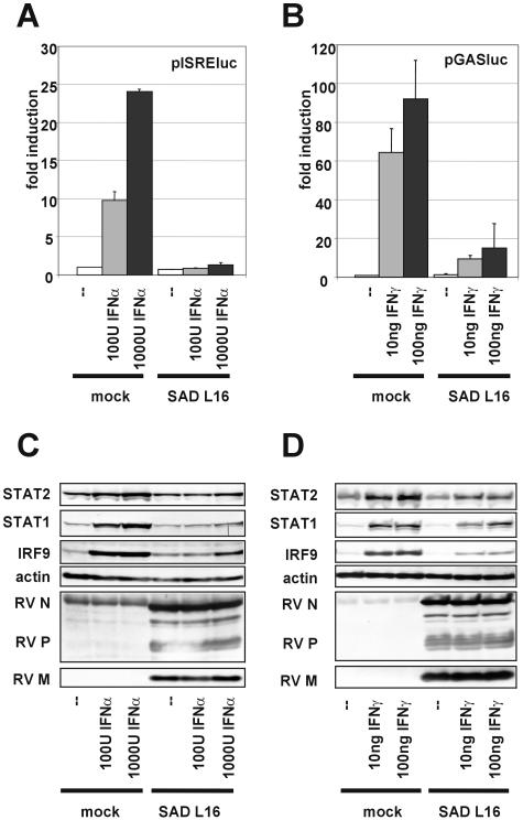
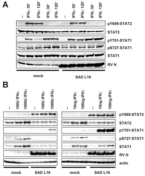
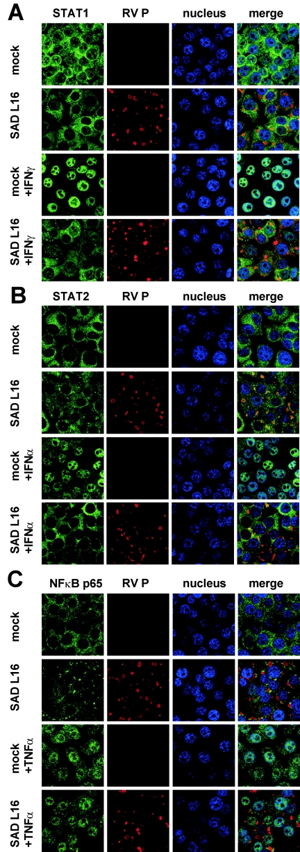
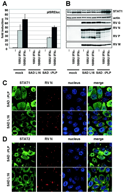
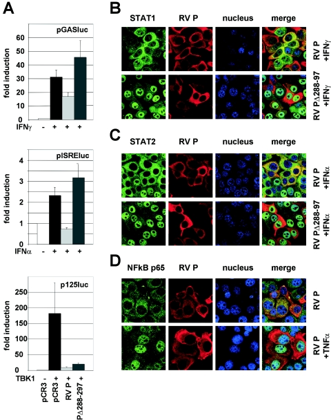
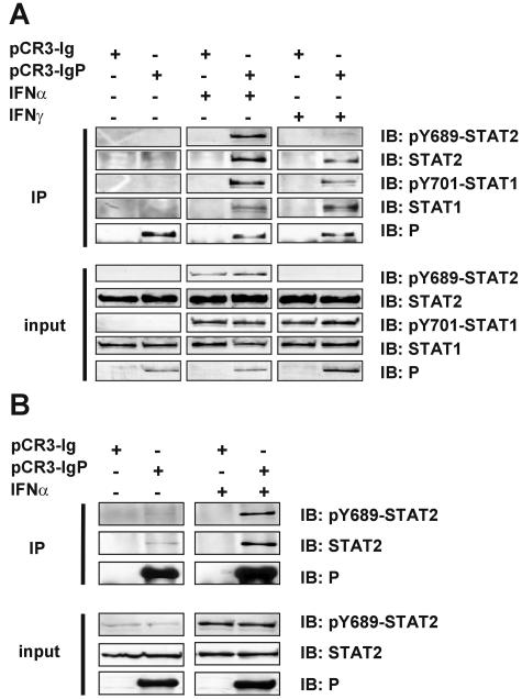
Rabies virus (RV) phosphoprotein P is an interferon (IFN) antagonist counteracting transcriptional activation of type I IFN (K. Brzózka, S. Finke, and K. K. Conzelmann, J. Virol 79:7673-7681, 2005). We here show that RV P in addition is responsible for preventing IFN-alpha/beta- and IFN-gamma-stimulated JAK-STAT signaling in RV-infected cells by the retention of activated STATs in the cytoplasm. Expression of IFN-stimulated response element- and gamma-activated sequence-controlled genes was severely impaired in cells infected with RV SAD L16 or in cells expressing RV P protein from transfected plasmids. In contrast, a recombinant RV expressing small amounts of P had lost the ability to interfere with JAK-STAT signaling. IFN-mediated tyrosine phosphorylation of STAT1 and STAT2 was not impaired in RV P-expressing cells; rather, a defect in STAT recycling was suggested by distinct accumulation of tyrosine-phosphorylated STATs in cell extracts. In the presence of P, activated STAT1 and STAT2 were unable to accumulate in the nucleus. Notably, STAT1 and STAT2 were coprecipitated with RV P only from extracts of cells previously stimulated with IFN-alpha or IFN-gamma, whereas in nonstimulated cells no association of P with STATs was observed. This conditional, IFN activation-dependent binding of tyrosine-phosphorylated STATs by RV P is unique for a viral IFN antagonist. The 10 C-terminal residues of P are required for counteracting JAK-STAT signaling but not for inhibition of transcriptional activation of IFN-beta, thus demonstrating two independent functions of RV P in counteracting the host's IFN response.


Figures

FIG. 1.
FIG. 1.
RV inhibits induction of ISGs by IFN-α and IFN-γ. (A and B) HEp-2 cells were infected with RV SAD L16 (MOI of 1), transfected with pISRE-luc (A) or pGAS-luc (B) at 18 h postinfection, and IFN-α or IFN-γ was added at 6 h posttransfection. Twenty-four hours later firefly luciferase activities were measured and corrected to the Renilla luciferase activity from a cotransfected control plasmid to normalize for variations in the transfection efficiency. Transfection experiments were repeated at least three times and averages and error bars are shown. (C and D) Expression of individual ISGs STAT1, STAT2, and IRF9 was analyzed by Western blot analysis of extracts from HEp-2 cells infected at an MOI of 1 for 24 h and a subsequent 24-h stimulation with IFN-α (C) or IFN-γ (D). In contrast to mock-infected cells, RV-infected cells do not upregulate ISRE- or GAS-controlled luciferase or STAT1, STAT2, and IRF9 upon IFN stimulation.
FIG. 2.
FIG. 2.
RV infection does not prevent tyrosine phosphorylation of STAT1 and STAT2 (A) but leads to accumulation of tyrosine-phosphorylated STAT1 and STAT2 over time (B). Mock-infected and RV-infected HEp-2 cells (MOI of 1) were stimulated at 24 h postinfection with IFN-α or IFN-γ. After 30 or 120 min (A) cell extracts were processed for Western blotting and probed with phospho-specific antibodies as indicated. Tyrosine phosphorylation of STAT2 (pY689-STAT2) and STAT1 (pY701-STAT1) was similar in mock- and RV-infected IFN-stimulated cells, whereas serine phosphorylation of STAT1 (pS727-STAT1) by IFN was more effective in mock-infected cells. Tyrosine-phosphorylated STAT1 and STAT2 accumulate. As shown in panel B, at later stages after IFN stimulation (24 h poststimulation), abundant amounts of tyrosine-phosphorylated STATs are still detectable only in RV-infected cells but not in mock-infected cells, suggesting a defect in STAT recycling.
FIG. 3.
FIG. 3.
RV infection prevents IFN-mediated translocation of STAT1 (A) and STAT2 (B) to the nucleus. RV-infected (MOI of 1) and mock-infected Hep-2 cells were stimulated with IFN-γ or IFN-α for 45 min at 24 h postinfection, fixed with 3% paraformaldehyde, and stained with STAT1 or STAT2 antibodies as indicated. The import of both STATs on IFN-treated cells is prevented in RV-infected cells, whereas import of NF-κB p65 on TNF-α-treated cells is not (C). Nuclei of cells were visualized by staining with TO-PRO-3 dye; RV P was visualized by using P-specific polyclonal mouse serum.
FIG. 4.
FIG. 4.
SAD ΔPLP fails in inhibiting JAK-STAT signaling. (A) BSR-T7/5 cells infected at an MOI of 3 with wt RV SAD L16 and a mutant expressing low levels of P (SAD ΔPLP) were transfected with pISRE-luc and were stimulated with IFN-α 18 h postinfection. In contrast to wt RV, SAD ΔPLP is not able to abolished luciferase activities. (B) BSR T7/5 cells infected at an MOI of 1 with RV SAD L16 and SAD ΔPLP were treated with IFN-α at 24 h postinfection. Cell lysates were prepared 24 h after stimulation (48 h postinfection) and were analyzed by Western blotting. Accumulation of viral proteins in SAD ΔPLP-infected cells was decreased after IFN induction. (C) HEp-2 cells infected with SAD L16 or SAD ΔPLP at an MOI of 1 were immunostained for STAT1 and STAT2 24 h postinfection as described in the legend of Fig. 3. In contrast to wt SAD L16, SAD ΔPLP-infected HEp-2 cells allow nuclear accumulation of both STAT1 and STAT2. RV N was visualized using N-specific mouse monoclonal antibodies.
FIG. 5.
FIG. 5.
Expression of P alone prevents STAT signaling. (A) pISRE-luc or pGAS-luc reporter constructs were cotransfected with plasmids encoding wt P (pCR3-RVP), a C-terminally truncated protein, PΔ288-297 (pCR3-RVPΔ288-297), or with empty vector (pCR3) into HEp-2 cells. After 24 h, IFN-α or IFN-γ was added, and luciferase activities were analyzed after an additional 24 h (top and middle panels). A reporter gene construct controlled by the IFN-β promoter (p125luc) was cotransfected with RV P and TBK-1 expression plasmids into HEK 293 cells as indicated. Luciferase activities were determined at 48 h posttransfection (lower panel). The average of at least two independent experiments is shown with error bars. (B to D) RV P prevents nuclear accumulation of STATs. HEp-2 cells were transfected with the indicated expression constructs. After 24 h cells were stimulated for 45 min and were subsequently stained for STAT1 (B), STAT2 (C), and NF-κB p65 (D) as described in the legend of Fig. 3. In contrast to PΔ288-297, expression of wt P prevents nuclear import of STAT1 and STAT2 by IFN but not of NF-κB p65 by TNF-α.
FIG. 6.
FIG. 6.
RV P interacts with STATs in an IFN-dependent manner. (A) HEK 293 cells were transfected with constructs encoding Ig-tagged P (pCR3-IgP) or the Ig moiety alone (pCR3-Ig). Precipitation was performed using Sepharose A beads binding the Ig tag at 48 h postinfection from extracts of cells that were treated for 45 min with 1,000 U/ml IFN-α or 100 ng/ml IFN-γ or were not treated. Precipitates (IP) and 3% of cell extracts (input) were analyzed by Western blotting with the indicated antibodies. Only from IFN-treated cells were STATs coprecipitated with Ig-P. (B) The experiment shown in panel A was performed with U3A cells that lack STAT1. STAT2 is precipitated from IFN-treated cells independent of STAT1.

Similar articles

Cited by

References

    1. Aaronson, D. S., and C. M. Horvath. 2002. A road map for those who don't know JAK-STAT. Science 296:1653-1655. - PubMed
    1. Akira, S., and K. Takeda. 2004. Toll-like receptor signalling. Nat. Rev. Immunol. 4:499-511. - PubMed
    1. Andrejeva, J., K. S. Childs, D. F. Young, T. S. Carlos, N. Stock, S. Goodbourn, and R. E. Randall. 2004. The V proteins of paramyxoviruses bind the IFN-inducible RNA helicase, mda-5, and inhibit its activation of the IFN-beta promoter. Proc. Natl. Acad. Sci. USA 101:17264-17269. - PMC - PubMed
    1. Bowie, A. G., and I. R. Haga. 2005. The role of Toll-like receptors in the host response to viruses. Mol. Immunol. 42:859-867. - PubMed
    1. Brzozka, K., S. Finke, and K. K. Conzelmann. 2005. Identification of the rabies virus alpha/beta interferon antagonist: phosphoprotein P interferes with phosphorylation of interferon regulatory factor 3. J. Virol. 79:7673-7681. - PMC - PubMed

Publication types