. 2006 Mar;80(6):2694-704.
doi: 10.1128/JVI.80.6.2694-2704.2006.

Annexin 2: a novel human immunodeficiency virus type 1 Gag binding protein involved in replication in monocyte-derived macrophages

Affiliations

Annexin 2: a novel human immunodeficiency virus type 1 Gag binding protein involved in replication in monocyte-derived macrophages

Elena V Ryzhova et al. J Virol. 2006 Mar.

Abstract

Human immunodeficiency virus (HIV) replication in the major natural target cells, CD4+ T lymphocytes and macrophages, is parallel in many aspects of the virus life cycle. However, it differs as to viral assembly and budding, which take place on plasma membranes in T cells and on endosomal membranes in macrophages. It has been postulated that cell type-specific host factors may aid in directing viral assembly to distinct destinations. In this study we defined annexin 2 (Anx2) as a novel HIV Gag binding partner in macrophages. Anx2-Gag binding was confined to productively infected macrophages and was not detected in quiescently infected monocyte-derived macrophages (MDM) in which an HIV replication block was mapped to the late stages of the viral life cycle (A. V. Albright, R. M. Vos, and F. Gonzalez-Scarano, Virology 325:328-339, 2004). We demonstrate that the Anx2-Gag interaction likely occurs at the limiting membranes of late endosomes/multivesicular bodies and that Anx2 depletion is associated with a significant decline in the infectivity of released virions; this coincided with incomplete Gag processing and inefficient incorporation of CD63. Cumulatively, our data suggest that Anx2 is essential for the proper assembly of HIV in MDM.


Figures

FIG. 1.
FIG. 1.
Anx2 interacts with p55Gag in MDM, transfected 293T cells, and cell-free systems. (a) Anti-Anx2 blot showing that Anx2 coprecipitates with Gag from MDM. Gag was immunoprecipitated from infected and uninfected MDM lysates with p24 rabbit polyclonal antiserum. Anx2 is present in the protein G-bound fraction from infected MDM only. (b) Anti-Anx2 blot showing Anx2-Gag coprecipitation from cotransfected 293T cells. Lysates from 293T cells transfected with a plasmid encoding the YU-2 proviral genome and Anx2-His or Anx2-His alone were precipitated with a rabbit polyclonal antiserum against Gag/p17. Anx2 coprecipitates with Gag in the doubly transfected cells. (c and d) Anti-Gag and anti-Anx2 Western blots. Purified Anx2-His protein precipitated on a nickel matrix after incubation with purified p55Gag specifically coprecipitated p55 Gag (c, left panel). When His-tagged amyloid precursor protein was substituted for Anx2, no precipitation of Gag was detected (c, right panel). Incubation in the presence of 100 μM calcium enhanced Anx2-p55Gag binding (d, left panel) without increasing nonspecific binding (d, right panel). Abbreviations: IgG, HC, immunoglobulin G heavy chain; IgG, HL, IgG light chain. B, bound fraction; w, wash. IP, immunoprecipitation; APP, amyloid precursor protein. Molecular masses: Anx2, 36 kDa; Gag/p55, 55 kDa; IgG, HC, 50 kDa; IgG, LH, 25 kDa.
FIG. 2.
FIG. 2.
Reciprocal distribution of Anx2, Gag, and CD63 in macrophages. Infected macrophages were fixed at day 4 after infection and immunostained for Anx2 and Gag (A) or for CD63 and Gag (B); uninfected macrophages were immunostained for Anx2 and CD63 (C) as specified in Materials and Methods. Anx2 and CD63 are shown in red; Gag is shown in green. The insets show additional 2× magnification of intracellular vesicles. Bar, 10 μm.
FIG. 3.
FIG. 3.
Depletion of Anx2 in MDM results in decreased viral replication. MDM were treated with Anx2-specific siRNA (siAnx520) or with nontargeting control siRNA (siAnxScr) and infected with HIV-1YU2 at day 3 after the treatment. Culture medium was collected and cells were lysed at 2, 4, and 7 days postinfection (p.i.). (A) The Anx2 level in cell lysates was analyzed by Western blotting at the indicated time points after infection. After Anx2 detection, membranes were stripped and reblotted for GAPDH as a control for total protein loads. (B and C) Release of p24Gag (B) and infectious units (C) in culture medium were measured using p24Gag ELISA and MAGI assay, respectively. (D) Intracellular Gag expression was monitored by Western blotting at days 2, 4, and 7 after infection. This experiment is representative of data from three independent donors.
FIG. 4.
FIG. 4.
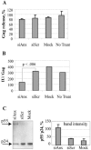
Depletion of Anx2 results in aberrant virus production. MDM were treated with siRNA as indicated and infected with HIV-1YU2 at day 3 after the treatment. Supernatants and cell lysates were collected at day 4 after infection. (A) Gag was measured in the lysates and cell-free supernatants by using p24 ELISA, which recognizes both p24Gag and unprocessed p55Gag. The efficiency of Gag release was determined as a percentage of intracellular Gag with respect to the extracellular Gag. (B) Infectious units in the cell-free supernatants were measured using MAGI assay and normalized by extracellular Gag measured in panel A. A decrease in the IU/p24 ratio indicates the production of replication-defective virions in the cells treated with siRNA specific for Anx2. (C) Cell-free supernatants were lysed and used in immunoprecipitation with anti-Gag rabbit polyclonal serum followed by Western blotting with anti-p24 mouse monoclonal antibody. The right panel shows the ratio of p24 band intensity to that of p55 measured using NIH ImageJ software. Error bars indicate standard deviations.
FIG. 5.
FIG. 5.
Anx2 depletion impairs virus maturation. (A) Infected macrophages stained with a monoclonal antibody against the mature form of the p17 matrix protein (red) or with anti-Gag polyclonal serum (green) at day 4 after infection. The merged panel shows that the p17 signal for mature HIV particles is collocated with regions of Gag accumulation, indicating sites of virus assembly and maturation (yellow). (B) HIV-1YU-2-infected, Anx2 siRNA-treated macrophages. Despite efficient p55Gag production visualized by the diffuse green staining, neither discrete sites of Gag accumulation nor mature virions were observed. (C) Efficiency of virion maturation was quantified as a percentage of infected cells positive for maturation marker (p55Gag+/p17Gag+) with respect to the total number of infected cells (p55Gag+). Data from two independent donors are shown; the range of infected cells was 53 to 100 per condition.
FIG. 6.
FIG. 6.
Anx2 depletion inhibits virus entry into MVBs. (A) Electron microscopy analysis of MDM transfected with scrambled siRNA and infected with HIV-1JAGO for 5 days. The inset shows a detailed view of particles with lentiviral morphology in intracellular vesicles resembling MVBs. (B) Cryosection of MDM treated with siAnx520 and infected as for panel A. No mature HIV particles either in intracellular vesicles or at the plasma membrane were detected upon Anx2 downregulation. (C) Virus capture with anti-CD63 antibody. Cell-free supernatants of MDM treated with siRNA and infected with HIV-1YU-2 were precipitated by Zysorbin cells with or without anti-CD63 antibody. Captured p24 was calculated by subtraction of p24 output remaining unprecipitated in the presence of anti-CD63 antibody from that in the supernatants precipitated without antibody. Efficiency of capture was expressed as the percentage of the value for the no-antibody control. Error bars indicate standard deviations.
FIG. 7.
FIG. 7.
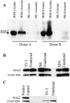
Annexin 2 is expressed in MDM but not in PBLs independently of infection status. (A) Donor-matched (donor A or donor B) uninfected PBLs and MDM in both the Triton-X-soluble and -insoluble fractions were probed for Anx2 protein expression. Anx2 was detected only in MDM in both fractions. (B) MDM were infected with various HIV-1 R5 strains, (YU-2, BaL, or ADA); lysates were taken 4 to 8 days postinfection and probed for Anx2 expression. Fifteen micrograms of total protein was loaded per well. (C) HIV-1YU-2-infected and uninfected stimulated PBLs were probed for Anx2 expression by Western blotting. Twenty micrograms of total protein was loaded per well.

Similar articles

Cited by

References

    1. Abdurahman, S., S. Hoglund, L. Goobar-Larsson, and A. Vahlne. 2004. Selected amino acid substitutions in the C-terminal region of human immunodeficiency virus type 1 capsid protein affect virus assembly and release. J. Gen. Virol. 85:2903-2913. - PubMed
    1. Adamson, C. S., A. Davies, Y. Soneoka, M. Nermut, K. Mitrophanous, and I. M. Jones. 2003. A block in virus-like particle maturation following assembly of murine leukaemia virus in insect cells. Virology 314:488-496. - PubMed
    1. Albright, A. V., J. T. Shieh, M. J. O'Connor, and Gonzalez-Scarano, F. 2000. Characterization of cultured microglia that can be infected by HIV-1. J. Neurovirol. 6(Suppl. 1):S53-S60. - PubMed
    1. Albright, A. V., R. M. Vos, and F. Gonzalez-Scarano. 2004. Low-level HIV replication in mixed glial cultures is associated with alterations in the processing of p55(Gag). Virology 325:328-339. - PubMed
    1. Brownstein, C., A. B. Deora, A. T. Jacovina, R. Weintraub, M. Gertler, K. M. Khan, D. J. Falcone, and K. A. Hajjar. 2004. Annexin II mediates plasminogen-dependent matrix invasion by human monocytes: enhanced expression by macrophages. Blood 103:317-324. - PubMed

Publication types