. 2006 Mar;80(6):2760-70.
doi: 10.1128/JVI.80.6.2760-2770.2006.

Beta1 integrin mediates internalization of mammalian reovirus

Affiliations

Beta1 integrin mediates internalization of mammalian reovirus

Melissa S Maginnis et al. J Virol. 2006 Mar.

Abstract

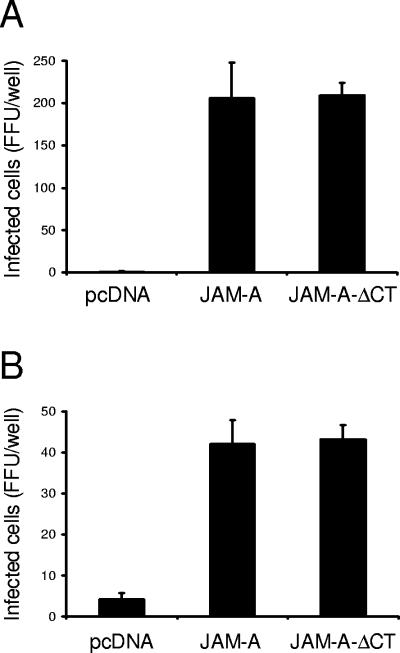
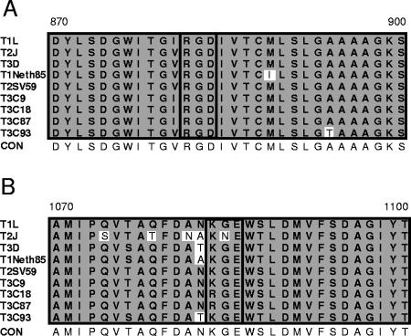
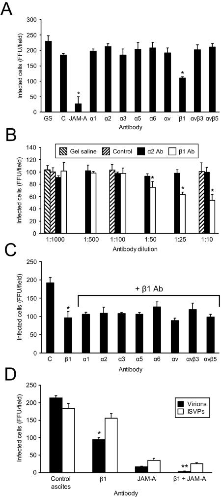
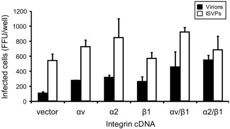
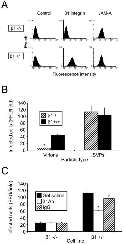
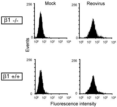
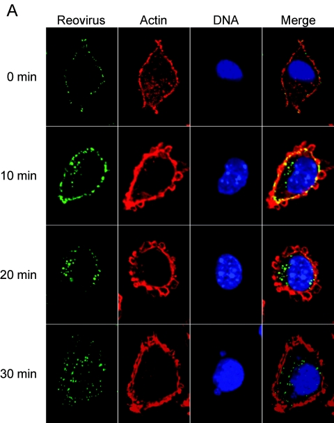
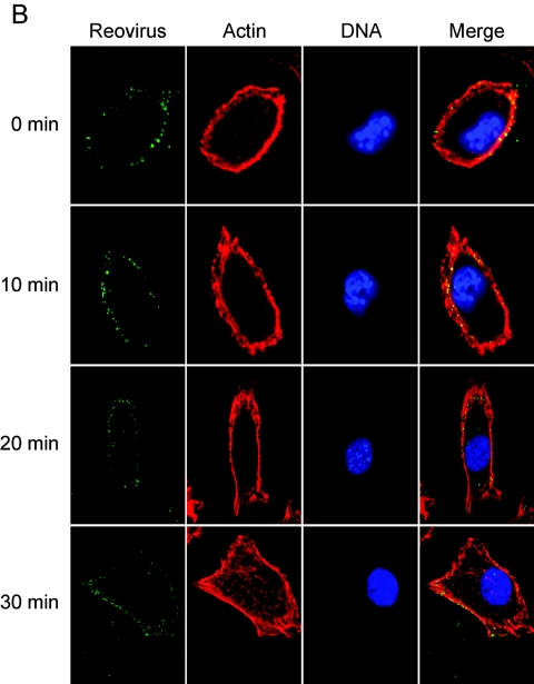
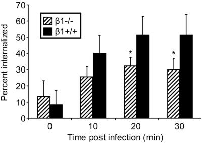
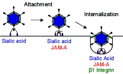
Reovirus infection is initiated by interactions between the attachment protein sigma1 and cell surface carbohydrate and junctional adhesion molecule A (JAM-A). Expression of a JAM-A mutant lacking a cytoplasmic tail in nonpermissive cells conferred full susceptibility to reovirus infection, suggesting that cell surface molecules other than JAM-A mediate viral internalization following attachment. The presence of integrin-binding sequences in reovirus outer capsid protein lambda2, which serves as the structural base for sigma1, suggests that integrins mediate reovirus endocytosis. A beta1 integrin-specific antibody, but not antibodies specific for other integrin subunits, inhibited reovirus infection of HeLa cells. Expression of a beta1 integrin cDNA, along with a cDNA encoding JAM-A, in nonpermissive chicken embryo fibroblasts conferred susceptibility to reovirus infection. Infectivity of reovirus was significantly reduced in beta1-deficient mouse embryonic stem cells in comparison to isogenic cells expressing beta1. However, reovirus bound equivalently to cells that differed in levels of beta1 expression, suggesting that beta1 integrins are involved in a postattachment entry step. Concordantly, uptake of reovirus virions into beta1-deficient cells was substantially diminished in comparison to viral uptake into beta1-expressing cells. These data provide evidence that beta1 integrin facilitates reovirus internalization and suggest that viral entry occurs by interactions of reovirus virions with independent attachment and entry receptors on the cell surface.


Figures

FIG. 1.
FIG. 1.
JAM-A cytoplasmic tail is not required for reovirus infection. CHO cells were transiently transfected with empty vector or plasmids encoding JAM-A or JAM-A-ΔCT. Following incubation for 24 h to permit receptor expression, cells were adsorbed with reovirus strains T1L (A) or T3D (B) at an MOI of 0.1 FFU per cell at room temperature for 1 h. Cells were washed with PBS, incubated in complete medium at 37°C for 20 h, and stained by indirect immunofluorescence. Infected cells were quantified by counting cells exhibiting cytoplasmic staining in entire wells for triplicate experiments. The results are expressed as the mean FFU per well for triplicate samples. Error bars indicate standard deviations. CHO cells support a low level of infection by type 3 reovirus in the absence of JAM-A, likely attributable to the expression of sialic acid (16, 34).
FIG. 2.
FIG. 2.
Reovirus outer capsid protein λ2 contains integrin-binding sequences. Alignment of deduced amino acid sequences of the reovirus λ2 protein for the indicated strains. Amino acid residues are designated by the single-letter code. Amino acid positions are indicated above the first and last letters. Integrin-binding RGD (A) and KGE (B) motifs are highlighted by a black box. Nonconserved sequences are shown in unshaded boxes. CON, consensus sequence.
FIG. 3.
FIG. 3.
β1 integrin antibody reduces reovirus infection of HeLa cells. HeLa cells were treated with gel saline (GS), control ascites (C), JAM-A-specific MAb J10.4, or antibodies specific for the α and β integrins shown (20 μg per ml or as diluted ascites) (A), gel saline, control ascites (Control), α2-specific MAb AA10, or β1-specific MAb DE9 (at the indicated dilutions) (B), antibodies specific for the α and β integrins shown in the presence of β1-specific MAb DE9 (1:10) (C), control ascites, β1-specific MAb DE9, JAM-A-specific MAb J10.4, or JAM-A-specific MAb J10.4 in combination with β1-specific MAb DE9 (D), and incubated at room temperature for 1 h. Antibody-treated cells were infected with virions or ISVPs of T1L at an MOI of 0.1 FFU per cell at 4°C for 30 min. Cells were washed with PBS, incubated in complete medium at 37°C for 16 h, and stained by indirect immunofluorescence. Infected cells were quantified by counting cells exhibiting cytoplasmic staining in three fields of view for triplicate samples. The results are expressed as the mean FFU per field for triplicate experiments. Error bars indicate standard deviations. *, P < 0.05 in comparison to the control; **, P < 0.05 in comparison to HeLa cells treated with JAM-A-specific MAb J10.4 alone.
FIG. 4.
FIG. 4.
β1 integrin expression enhances reovirus infection of CEFs. CEFs were transiently transfected with JAM-A-encoding plasmid alone (vector) or in combination with plasmids encoding the integrin subunits shown. Following 24 h to allow receptor expression, transfected cells were adsorbed with T1L virions or ISVPs at an MOI of 1 FFU per cell at room temperature for 1 h. Cells were washed with PBS, incubated in complete medium at 37°C for 20 h, and stained by indirect immunofluorescence. Infected cells were quantified by counting cells exhibiting cytoplasmic staining in entire wells for duplicate samples. The results are expressed as the mean FFU per well for duplicate experiments. Error bars indicate the range of data. Shown is a representative experiment of three independent experiments performed.
FIG. 5.
FIG. 5.
Cells deficient in β1 integrin are less permissive for reovirus infection. (A) GD25 (β1−/−) and GD25β1A (β1+/+) cells were detached from plates with 20 mM EDTA, washed, and incubated with antibodies specific for either murine β1 integrin or murine JAM-A. Cell surface expression of these molecules was detected by flow cytometry. Data are expressed as fluorescence intensity. GD25 and GD25β1A cells were untreated (B) or pretreated with β1-specific MAb CD29 (β1 Ab) or a hamster isotype-matched control MAb (IgG) at room temperature for 1 h (C), adsorbed with virions or ISVPs of T1L at an MOI of 0.1 FFU per cell, and incubated at 4°C for 30 min. Cells were washed with PBS, incubated in complete medium at 37°C for 20 h, and stained by indirect immunofluorescence. Infected cells were quantified by counting cells exhibiting cytoplasmic staining in five fields of view for duplicate samples. The results are expressed as the mean FFU per field for triplicate experiments. *, P < 0.05 in comparison to the control.
FIG. 6.
FIG. 6.
Reovirus exhibits equivalent binding to β1−/− and β1+/+ cells. GD25 (β1−/−) and GD25β1A (β1+/+) cells were incubated with either PBS (mock) or 2 × 10¹¹ FITC-labeled T1L virions at 4°C for 1 h and analyzed by flow cytometry to assess reovirus binding to the cell surface. The results are expressed as fluorescence intensity.
FIG.7.
FIG.7.
β1 integrin enhances reovirus entry into cells. (A) GD25β1A (β1+/+) and (B) GD25 (β1−/−) cells were chilled, adsorbed with T1L virions, and incubated at 4°C for 1 h. Nonadherent virus was removed, warm medium was added, and cells were incubated at 37°C for the times shown. Cells were fixed, stained for reovirus (green), actin (red), and DNA (blue), and imaged using confocal immunofluorescence microscopy. Representative digital fluorescence images of the same field are shown in each row.
FIG.7.
FIG.7.
β1 integrin enhances reovirus entry into cells. (A) GD25β1A (β1+/+) and (B) GD25 (β1−/−) cells were chilled, adsorbed with T1L virions, and incubated at 4°C for 1 h. Nonadherent virus was removed, warm medium was added, and cells were incubated at 37°C for the times shown. Cells were fixed, stained for reovirus (green), actin (red), and DNA (blue), and imaged using confocal immunofluorescence microscopy. Representative digital fluorescence images of the same field are shown in each row.
FIG. 8.
FIG. 8.
Quantification of reovirus internalization into β1−/− and β1+/+ cells. Viral internalization was quantitated by enumerating fluorescent particles localized at the cell periphery and particles internalized into the cytoplasm to determine the total number of fluorescent particles per cell. The results are expressed as mean percent internalization (internalized fluorescent particles/total number of fluorescent particles per cell) for 10 cells for each time point. *, P < 0.05 in comparison to β1+/+ cells.
FIG. 9.
FIG. 9.
Receptors for reovirus attachment and cell entry. Reovirus initially engages cells by low-affinity interactions with carbohydrate. For type 3 reovirus strains, this carbohydrate is sialic acid. Reovirus-carbohydrate interactions are followed by high-affinity binding to JAM-A, which positions the virus on the cell surface for subsequent interactions with β1 integrin to trigger viral endocytosis.

Similar articles

Cited by

References

    1. Akula, S. M., N. P. Pramod, F. Z. Wang, and B. Chandran. 2002. Integrin α3b1 (CD 49c/29) is a cellular receptor for Kaposi's sarcoma-associated herpesvirus (KSHV/HHV-8) entry into the target cells. Cell 108:407-419. - PubMed
    1. Baer, G. S., and T. S. Dermody. 1997. Mutations in reovirus outer capsid protein σ3 selected during persistent infections of L cells confer resistance to protease inhibitor E64. J. Virol. 71:4921-4928. - PMC - PubMed
    1. Baer, G. S., D. H. Ebert, C. J. Chung, A. H. Erickson, and T. S. Dermody. 1999. Mutant cells selected during persistent reovirus infection do not express mature cathepsin L and do not support reovirus disassembly. J. Virol. 73:9532-9543. - PMC - PubMed
    1. Barton, E. S., J. L. Connolly, J. C. Forrest, J. D. Chappell, and T. S. Dermody. 2001. Utilization of sialic acid as a coreceptor enhances reovirus attachment by multistep adhesion strengthening. J. Biol. Chem. 276:2200-2211. - PubMed
    1. Barton, E. S., J. C. Forrest, J. L. Connolly, J. D. Chappell, Y. Liu, F. Schnell, A. Nusrat, C. A. Parkos, and T. S. Dermody. 2001. Junction adhesion molecule is a receptor for reovirus. Cell 104:441-451. - PubMed

Publication types

LinkOut - more resources