. 2006 Mar;80(6):2815-22.
doi: 10.1128/JVI.80.6.2815-2822.2006.

Detection of cell-cell fusion mediated by Ebola virus glycoproteins

Affiliations

Detection of cell-cell fusion mediated by Ebola virus glycoproteins

Séverine Bär et al. J Virol. 2006 Mar.

Abstract

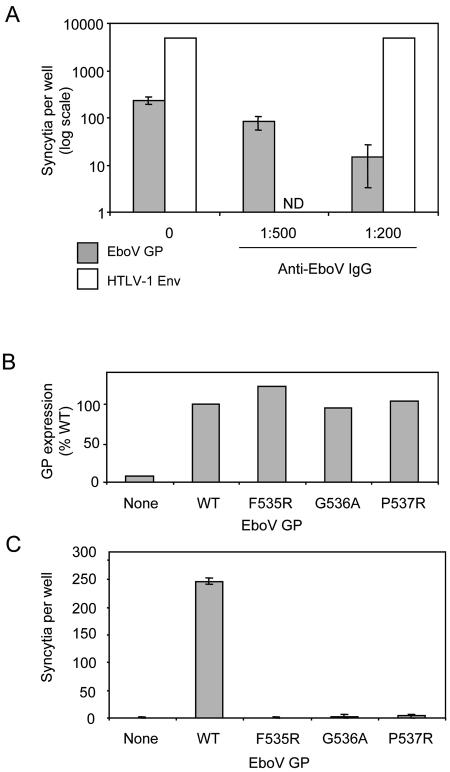
Ebola viruses (EboV) are enveloped RNA viruses infecting cells by a pH-dependent process mediated by viral glycoproteins (GP) involving endocytosis of virions and their routing into acidic endosomes. As with well-characterized pH-dependent viral entry proteins, in particular influenza virus hemagglutinin, it is thought that EboV GP require activation by low pH in order to mediate fusion of the viral envelope with the membrane of endosomes. However, it has not yet been possible to confirm the direct role of EboV GP in membrane fusion and the requirement for low-pH activation. It was in particular not possible to induce formation of syncytia by exposing cells expressing EboV GP to acidic medium. Here, we have used an assay based on the induction of a beta-galactosidase (lacZ) reporter gene in target cells to detect cytoplasmic exchanges, indicating membrane fusion, with cells expressing EboV GP (Zaire species). Acidic activation of GP-expressing cells was required for efficient fusion with target cells. The direct role of EboV GP in this process is indicated by its inhibition by anti-GP antibodies and by the lack of activity of mutant GP normally expressed at the cell surface but defective for virus entry. Fusion was not observed when target cells underwent acidic treatment, for example, when they were placed in coculture with GP-expressing cells before the activation step. This unexpected feature, possibly related to the nature of the EboV receptor, could explain the impossibility of inducing formation of syncytia among GP-expressing cells.


Figures

FIG. 1.
FIG. 1.
Detection of cell-cell fusion by activation of a lacZ reporter gene in target cells (A) or by diffusion of cytoplasmic fluorescent probes (B). (A) X-Gal staining after a 24-hour coculture of HeLa-CD4-LTRlacZ target cells with HeLa cells previously transfected with expression vectors for HIV-1 Tat (a), Tat and HIV-1 Env (b), or Tat and EboV GP (c) and exposed for 10 min to pH 5 medium (see Materials and Methods). Blue foci indicate fusion of target and Tat-positive cells. Magnification, ×4. (B) Confocal microscopy fields of cocultures of HeLa-CD4-LTRlacZ cells labeled with CMTMR (red fluorescence) and HeLa cells labeled with CTG (green fluorescence) after transfection with control vector (a), HIV-1 Env (b), or EboV GP (c) expression vectors. CTG-labeled cells were exposed to pH 5 medium for 10 min before a 24-h coculture in neutral medium with CMTMR-labeled cells. Pictures were taken at a ×63 magnification.
FIG. 2.
FIG. 2.
Quantification of cell-cell fusion mediated by EboV GP or HIV-1 Env. (A) Fusion with HeLa-CD4, HeLa, or COS target cell lines was detected as described in the legend to Fig. 1A by scoring blue foci after they were stained with X-Gal. Bars represent number of blue foci per well (six-well plates) in one of three experiments. ND, not done. (B) Effect of treatment of cells expressing EboV GP with acidic medium (pH 3 to 7) on fusion with HeLa-CD4-LTRlacZ target cells. The experiment was conducted as described in panel A, and bars represent means from three independent experiments. (C) Same experiment as that described in panel B, with COS-LTRlacZ target cells. One of two experiments with similar results is shown.
FIG. 3.
FIG. 3.
Inhibition of EboV GP-mediated cell-cell fusion by neutralizing antibodies (A) or mutations in the GP2 subunit (B and C). (A) Cocultures of HeLa-CD4-LTRlacZ cells with HeLa cells transfected with expression vectors for Tat and either EboV GP or HTLV-1 Env were performed as described in the legend to Fig. 2A, except for the presence of purified horse anti-GP IgGs at 1:500 or 1:200 dilution. Bars represent means of three replicates. (B) Effect of mutations in the putative fusion peptide of GP2 on the expression of EboV GP at the surface of transfected HeLa cells measured by flow cytometry after staining with a mouse anti-GP MAb and a secondary FITC-coupled antibody. Results are shown as percentages of GP-expressing cells relative to cells transfected with a WT GP expression vector. Data are from one of two representative experiments. (C) Effect of GP2 mutations on fusion with HeLa-CD4-LTRlacZ target cells. The experiment was performed as described in the legend to Fig. 1B. Results are the means from three representative experiments.
FIG. 4.
FIG. 4.
Effects of target cell treatment on EboV GP-mediated cell-cell fusion. (A) Cocultures of HeLa-CD4-LTRlacZ cells treated with proteinase K (1 h) or dextran sulfate (2 h) with HeLa cells expressing Tat and either EboV GP or VSV-G were performed and fusion events scored as described in the legend to Fig. 2A. (B) Effect of low-pH treatment (pH 5; 10 min) applied either to HeLa cells expressing EboV GP or VSV-G (effector cells) before coculture, to both effector and target cells before coculture, or after a 60-min contact of effector and target cells. In all cases, exposure to acidic medium was ended by a wash in neutral-pH medium. Bars represent numbers of blue foci per well after overnight coculture and staining with X-Gal. Two independent experiments (exp 1 and exp 2) are represented for EboV GP and one for VSV-G. The asterisk indicates massive fusion with large multinucleated syncytia (>10 nuclei). Scoring the number of blue foci underestimated fusion efficiency in this situation. (C) Efficiency of fusion after return of acid-treated target cells into neutral-pH medium. HeLa-CD4-LTRlacZ cells were detached from the plate, resuspended in pH 5 medium for 10 min at room temperature (RT), and then returned to culture medium at 37°C. At different times, a fraction of these cells was added to acid-treated HeLa cells expressing either EboV GP or VSV-G, and fusion was monitored as described in the legend to Fig. 2A. Numbers of fusion events per well are shown as percentages relative to untreated target cells (means from three independent experiments).
FIG. 5.
FIG. 5.
Kinetics of cell fusion efficiency after low-pH activation of EboV GP and VSV-G. The experiment was conducted as described in the legend to Fig. 2A except that HeLa cells expressing Tat and EboV GP or VSV-G were left in neutral-pH medium from 15 to 120 min after the acid activation step and before coculture in neutral medium with HeLa-CD4-LTRlacZ cells. Numbers of fusion events per well are shown as percentages relative to cocultures initiated immediately after low-pH exposure (means from three independent experiments).

References

    1. Bär, S., and M. Alizon. 2004. Role of the ectodomain of the gp41 transmembrane envelope protein of human immunodeficiency virus type 1 in late steps of the membrane fusion process. J. Virol. 78:811-820. - PMC - PubMed
    1. Chan, S. Y., C. J. Empig, F. J. Welte, R. F. Speck, A. Schmaljohn, J. F. Kreisberg, and M. A. Goldsmith. 2001. Folate receptor-alpha is a cofactor for cellular entry by Marburg and Ebola viruses. Cell 106:117-126. - PubMed
    1. Chan, S. Y., M. C. Ma, and M. A. Goldsmith. 2000. Differential induction of cellular detachment by envelope glycoproteins of Marburg and Ebola (Zaire) viruses. J. Gen. Virol. 81:2155-2159. - PubMed
    1. Chandran, K., N. J. Sullivan, U. Felbor, S. P. Whelan, and J. M. Cunningham. 2005. Endosomal proteolysis of the Ebola virus glycoprotein is necessary for infection. Science 308:1643-1645. - PMC - PubMed
    1. Chernomordik, L. V., E. Leikina, V. Frolov, P. Bronk, and J. Zimmerberg. 1997. An early stage of membrane fusion mediated by the low pH conformation of influenza hemagglutinin depends upon membrane lipids. J. Cell Biol. 136:81-93. - PMC - PubMed

Publication types