. 2006 Mar;80(6):2863-72.
doi: 10.1128/JVI.80.6.2863-2872.2006.

Innate immune responses to herpes simplex virus type 2 influence skin homing molecule expression by memory CD4+ lymphocytes

Affiliations

Innate immune responses to herpes simplex virus type 2 influence skin homing molecule expression by memory CD4+ lymphocytes

David M Koelle et al. J Virol. 2006 Mar.

Abstract

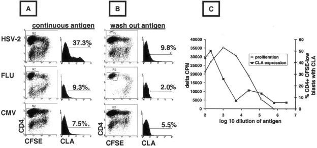
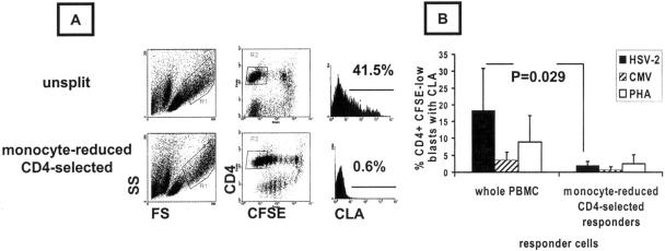
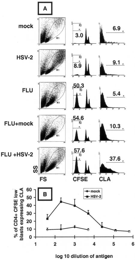
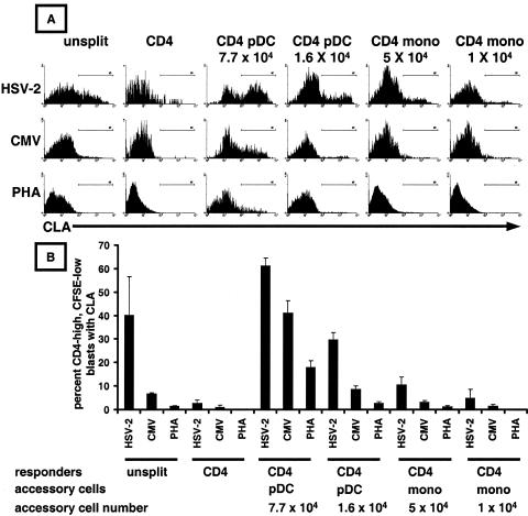
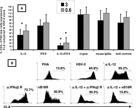
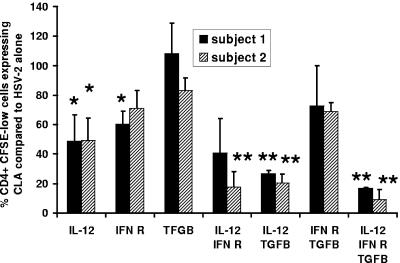
Herpes simplex virus (HSV) infections of humans are characterized by intermittent, lytic replication in epithelia. Circulating HSV-specific CD4 T cells express lower levels of preformed cutaneous lymphocyte-associated antigen (CLA), a skin-homing receptor, than do circulating HSV-specific CD8 T cells but, paradoxically, move into infected skin earlier than CD8 cells. Memory CD4 T cells develop strong and selective expression of CLA and E-selectin ligand while responding to HSV antigen in vitro. We now show that interleukin-12, type I interferon, and transforming growth factor beta are each involved in CLA expression by memory HSV type 2 (HSV-2)-specific CD4 T cells in peripheral blood mononuclear cells (PBMC). A reduction of the number of monocytes and dendritic cells from PBMC reduces CLA expression by HSV-2-responsive CD4 lymphoblasts, while their reintroduction restores this phenotype, identifying these cells as possible sources of CLA-promoting cytokines. Plasmacytoid dendritic cells are particularly potent inducers of CLA on HSV-reactive CD4 T cells. These observations are consistent with cooperation between innate and acquired immunity to promote a pattern of homing receptor expression that is physiologically appropriate for trafficking to infected tissues.


Figures

FIG. 1.
FIG. 1.
Dissociation of CLA induction and proliferation by HSV-2-reactive memory CD4 T cells. (A) PBMC were incubated continuously with antigen/mitogen and cultured for 5 days in 48-well plates. Large granular cells (gating not shown) that were CD4+ CFSElow (R2) were analyzed. Histograms and markers show the percent cells expressing CLA. (B) PBMC were pulsed with antigen/mitogen for 2 hours, washed, cultured, and analyzed as described above. The results shown in panels A and B are typical of five subjects. (C) PBMC from an HSV-1- and HSV-2-seropositive person were incubated with serial dilutions of whole HSV-1 or HSV-2 antigen. After 5 days, proliferation was measured in triplicate by [3H]thymidine incorporation. CLA expression was measured as described above. Results are typical of four subjects, and means are shown.
FIG. 2.
FIG. 2.
CD4-enriched, monocyte-depleted PBMC cultures proliferating in response to HSV-2 antigen have low CLA expression. (A) Representative unsplit or monocyte-depleted, CD4-enriched PBMC from HSV-2/CMV dually seropositive donors were stimulated for 5 days with viral antigen or PHA and analyzed for size and granularity (left) or CFSE dilution and CD4 expression (middle). Large CD4+ CFSElow cells, gated as shown, were analyzed for CLA, and the percentages of positive cells are shown. (B) Means and standard deviations for four subjects with matched whole PBMC and CD4-enriched, monocyte-depleted cultures analyzed in parallel. Each responder cell population and antigen was tested in duplicate in a 96-well format, and the means for individual subjects were used to calculate means (bars) and standard deviations (error bars).
FIG. 3.
FIG. 3.
HSV-2 antigen induces CLA expression on bystander proliferating CD4+ memory T cells. (A) PBMC from an HSV-seronegative, influenza-vaccinated subject were incubated for 5 days with the antigen(s) indicated at left. Total lymphocytes (resting and blasts) in the indicated R1 forward/side scatter window were gated for CD4hi (not shown). Influenza virus antigen led to more CFSElow cells, indicated numerically above the histograms in the middle column, than did HSV-2. CLA expression on the gated (R3) CD4+ CFSElow large granular cells is shown in the right column, and positive cell percentages are indicated. (B) Dose dependence of bystander CLA expression induced by UV-inactivated HSV-2 or mock antigen. Data are means and standard deviations of triplicate results.
FIG. 4.
FIG. 4.
Plasmacytoid dendritic cells induce CLA expression by memory CD4 T cells responding to HSV-2 antigen. (A) Representative histograms showing CLA expression after exposure of the indicated cell populations, gated for CD4+ CFSElow lymphoblasts, to viral antigens or PHA. Either the whole PBMC (left) or purified CD4+ T cells were labeled with CFSE. (B) Means and standard deviations of CLA expression levels from triplicate assays for a representative subject. Mono, monocytes.
FIG. 5.
FIG. 5.
(A) Blockade of IL-12 acts alone and synergistically with blockade of class I interferon to prevent CLA expression by HSV-2-reactive CD4 T cells. PBMC from five HSV-2-infected donors were CFSE labeled, and antibodies were added on day 0 with HSV-2 antigen (1:1,000). The dose of antibody (3 or 0.6 μg/ml) is indicated in the legend. On day 5, CFSElow CD4+ blasts were gated and examined for CLA expression. CLA expression in the absence of antibodies was set to 100% for each patient. Bars and error bars are means and standard deviations, respectively, for five subjects for antibody at 0.6 μg/ml (hatched) or 3.0 μg/ml (solid). Asterisks refer to significant (P < 0.05) reductions of CLA expression compared to the wells containing the relevant control antibody or antibodies. Control antibodies (right) led to no significant differences (P > 0.05) from the no-antibody control. (B) Effect of neutralizing antibodies and soluble recombinant vaccinia virus B18R protein on CLA expression in a representative HSV-2-seropositive subject. PBMC were incubated with PHA or HSV-2 antigen (1:1,000) in the absence or presence of the indicated neutralizing antibodies (0.6 μg/ml) or B18R (0.2 μg/ml). Gated blasts that were CFSElow CD4+ were analyzed for CLA expression. Data are percentages of cells positive for CLA.
FIG. 6.
FIG. 6.
Neutralization of TGF-β reduces CLA expression in combination with blockade of IL-12. PMBC from two HSV-2-infected donors (as indicated in the legend) were CFSE labeled, and antibodies (all used at 0.6 μg/ml) were added on day 0 in duplicate microtiter plate cultures. Cultures were analyzed on day 5, with CLA expression in the absence of antibodies set to 100%. Bars and error bars are means and standard deviations, respectively, of the percentages of CLA expression by CFSElow CD4+ blasts for two subjects relative to the no-antibody control. Single asterisks refer to significant (P < 0.05) reductions of CLA expression compared to the no-antibody control (100% on the y axis). Double asterisks refer to significant (P < 0.05) decreases of CLA expression compared to that with anti-IL-12 alone.

Similar articles

Cited by

References

    1. Alcami, A., J. A. Symons, and G. L. Smith. 2000. The vaccinia virus soluble alpha/beta interferon (IFN) receptor binds to the cell surface and protects cells from the antiviral effects of IFN. J. Virol. 74:11230-11239. - PMC - PubMed
    1. Allan, R. S., C. M. Smith, G. T. Belz, A. L. van Lint, L. M. Wakim, W. R. Heath, and F. R. Carbone. 2003. Epidermal viral immunity induced by CD8alpha+ dendritic cells but not by Langerhans cells. Science 301:1925-1928. - PubMed
    1. Ankel, H., D. F. Westra, S. Welling-Wester, and P. Lebon. 1998. Induction of interferon-alpha by glycoprotein D of herpes simplex virus: a possible role of chemokine receptors. Virology 251:317-326. - PubMed
    1. Armerding, D., and T. S. Kupper. 1999. Functional cutaneous lymphocyte antigen can be induced in essentially all peripheral blood T lymphocytes. Int. Arch. Allergy Immunol. 119:212-222. - PubMed
    1. Bangert, C., J. Friedl, G. Stary, G. Stingl, and T. Kopp. 2003. Immunopathologic features of allergic contact dermatitis in humans: participation of plasmacytoid dendritic cells in the pathogenesis of the disease? J. Investig. Dermatol. 121:1409-1418. - PubMed

Publication types

MeSH terms