. 2006 Mar;80(6):2884-93.
doi: 10.1128/JVI.80.6.2884-2893.2006.

A small loop in the capsid protein of Moloney murine leukemia virus controls assembly of spherical cores

Affiliations

A small loop in the capsid protein of Moloney murine leukemia virus controls assembly of spherical cores

Marcy R Auerbach et al. J Virol. 2006 Mar.

Abstract

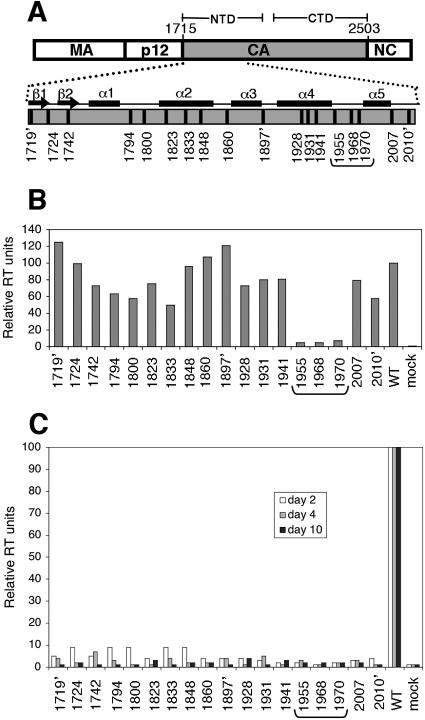
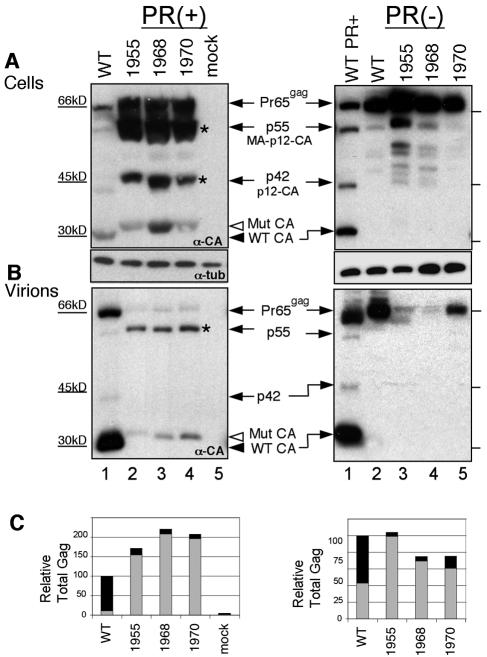
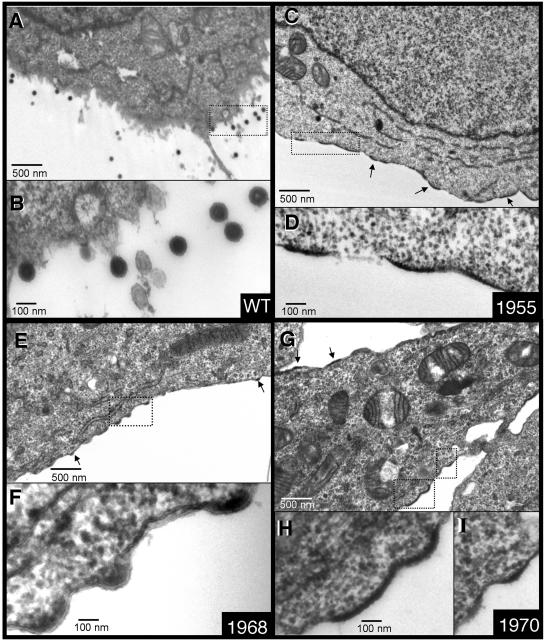
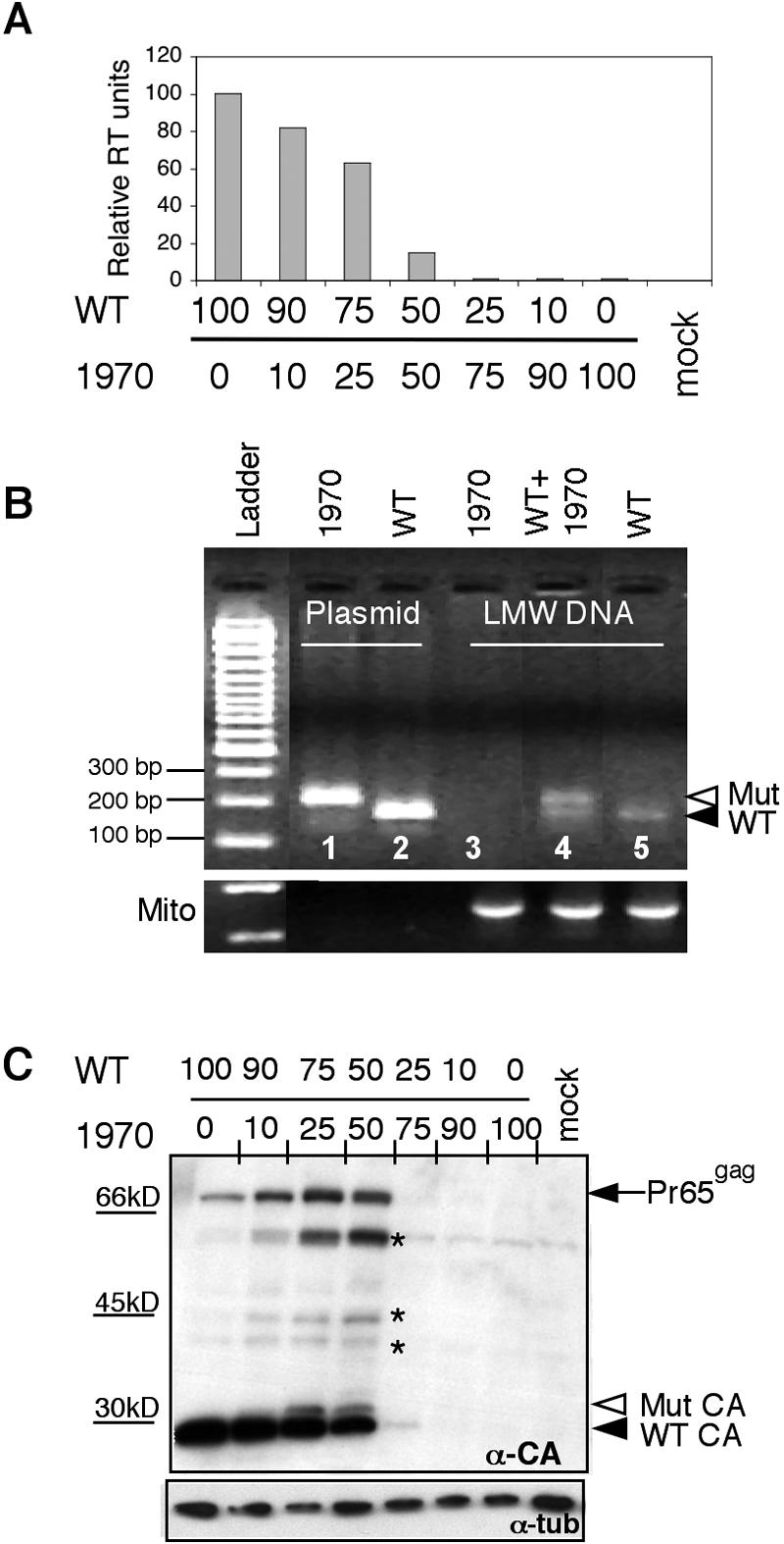
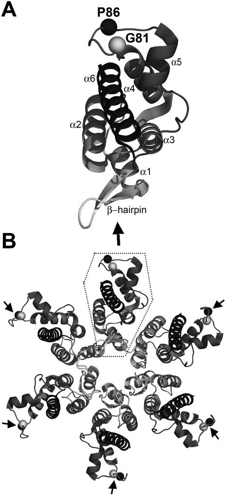
We report the identification of a novel domain in the Gag protein of Moloney murine leukemia virus (MoLV) that is important for the formation of spherical cores. Analysis of 18 insertional mutations in the N-terminal domain of the capsid protein (CA) identified 3 that were severely defective for viral assembly and release. Transmission electron microscopy of cells producing these mutants showed assembly of Gag proteins in large, flat or dome-shaped patches at the plasma membrane. Spherical cores were not formed, and viral particles were not released. This late assembly/release block was partially rescued by wild-type virus. All three mutations localized to the small loop between alpha-helices 4 and 5 of CA, analogous to the cyclophilin A-binding loop of human immunodeficiency virus type 1 CA. In the X-ray structure of the hexameric form of MLV CA, this loop is located at the periphery of the hexamer. The phenotypes of mutations in this loop suggest that formation of a planar lattice of Gag is unhindered by mutations in the loop. However, the lack of progression of these planar structures to spherical ones suggests that mutations in this loop may prevent formation of pentamers or of stable pentamer-hexamer interactions, which are essential for the formation of a closed, spherical core. This region in CA, focused to a few residues of a small loop, may offer a novel therapeutic target for retroviral diseases.


Figures

FIG. 1.
FIG. 1.
Insertional mutations in NTD of MoMLV CA. (A) MoMLV Gag polyprotein. CA polypeptide (nucleotides 1715 to 2503) is highlighted in gray, with 1′ being the first nucleotide of pNCA, the infectious MoMLV clone. The positions of the β-hairpin and α-helices 1 to 5 of CA are indicated (35). Locations of mutations are denoted by vertical bars. Mutants are designated by the nucleotide position at the 5′ end of the insertional mutation. The three mutants between α-helices 4 and 5 (marked by a bracket) are the focus of this study. Three mutants generated from a separate mutagenesis contained five-amino-acid insertions and are indicated with an apostrophe in all panels. (B) Viral production assayed by RT activity in supernatants of cells transfected with proviral DNAs. RT activity of WT virus was set to 100 (average of three experiments). (C) Infectivity of mutants as measured by virus spreading assay. Culture medium from cells transfected with mutant proviral DNAs was used to inoculate naïve Rat2 cells. Virions released at days 2, 4, and 10 after infection were measured by RT activity in harvested supernatants (white, gray, and black bars, respectively).
FIG. 2.
FIG. 2.
Gag proteins of release-defective mutants. (A) Viral protein expression in cells transfected with proviral DNA. Western blot analysis using anti-CA polyclonal serum detected CA and other Gag intermediates 48 h after transfection. The arrow indicates the position of the Gag precursor (Pr65gag) and intermediates. Open arrowhead, mutant CA protein; closed arrowhead, WT CA protein. Asterisks mark intermediates of Gag processing. The lower blot in panel A is the same blot and was probed with antitubulin antibody. Mutants in the context of an inactive viral protease are shown in the right panel. (B) Virions released into the medium following transfection with proviral DNA were concentrated and lysed, and the proteins were analyzed by Western blotting using anti-CA antibodies. (C) Particle release was defective for mutants 1955, 1968, and 1970. Gag proteins were quantified from blots. Gray bars represent intracellular Gag protein (from panel A, using lower exposures to ensure that signals were in the linear range for film), and black bars denote Gag released in pelleted virions (from panel B). The total amount of Gag protein in wild-type virus was set to 100%.
FIG. 3.
FIG. 3.
TEM images of cells transfected with proviral DNA from WT and CA mutant virus: WT (A and B), 1955 (C and D), 1968 (E and F), and 1970 (G to I). Each panel shows a field at ×20,000 magnification (A, C, E, and G). Areas within fields (marked by rectangles) are magnified 75,000-fold (B, D, F, H, and I). Arrows indicate Gag assemblies beneath the plasma membrane. Scale bars are as shown.
FIG. 4.
FIG. 4.
WT virus can rescue CA mutants. (A) Virions released upon introduction of a mixture of WT and mutant DNA into cells. On the left, cells were cotransfected with equal amounts of WT and 1970 DNA. On the right, increasing amounts of mutant DNA were used, keeping the total DNA amounts constant. Supernatants were analyzed for RT activity 48 h posttransfection. As a control, the right panel shows increasing amounts of nonviral DNA (expression plasmid for EGFP) added to WT DNA in cotransfections. (B) Western blot of pelleted virions released from cotransfected cells using anti-CA polyclonal serum. A short exposure allows mutant and WT CA proteins (open and closed arrowheads, respectively), differing in size by 12 amino acids, to be distinguished. EM images are of 293T cells cotransfected with WT and mutant proviral genomes at different ratios, as follows: (C and D) WT and 1970, 50:50; (E and F) WT and 1970, 25:75; (G and H) WT and 1970, 10:90. Arrows indicate Gag assembly at the membrane. Bar, 100 nm.
FIG. 5.
FIG. 5.
Mixed virions are infectious. (A) The ability of mixed virions to infect cells was assayed using the viral spreading assay. Culture medium was collected after cotransfection with WT and mutant proviral DNAs in the indicated ratios. Equal amounts of released virus were used to inoculate naïve Rat2 cells. RT activity was measured in supernatants harvested 36 h after inoculation. (B) PCR using low-molecular-weight DNA from infected Rat2 cells. PCR primers flanked the region containing the insertional mutations, producing a 243-bp product from WT viral DNA and 279 bp from the mutant. Template DNAs consisted of mutant and WT proviral plasmid DNA (controls in lanes 1 and 2) and low-molecular-weight DNA from Rat2 cells infected with mutant virions (lane 3), mixed virions (cotransfections were done with equal amounts of WT and 1970 proviral DNA; lane 4), and WT virions (lane 5). The WT resulted in a PCR product of the expected size (lower arrow), while 1970 alone resulted in no PCR product, since it did not make virions. Mixed virions resulted in a slower-migrating band corresponding to the mutant product and a faster one for WT (doublet). PCR products resulting from amplification of rat mitochondrial DNA (bottom gel in panel B) indicated that nearly equal amounts of template were used in the analysis. (C) Rat2 cell lysates were harvested 36 h after inoculation with mixed virions and analyzed by Western blotting using anti-CA polyclonal serum. Arrows indicate WT and mutant CA protein. The same blot reprobed with antitubulin antibodies is shown below to indicate equal loading of lanes on the gel.
FIG. 6.
FIG. 6.
Structure of the NTD of MLV CA. (A) Monomer of the NTD of MLV CA from published coordinates of the NTD hexamer. Spheres show the locations of the CA mutants in the loop between α-helices 4 and 5. Mutant 1955 is located between amino acids G81 and D82 (gray sphere); mutants 1968 and 1970 are located between P86 and T87 (black sphere). (B) Hexamer of the NTD of MLV CA (PDB, 1U7K) (28). The dotted box outlines the monomer illustrated in panel A. Drawings were made using PyMol (9).

References

    1. Accola, M. A., B. Strack, and H. G. Gottlinger. 2000. Efficient particle production by minimal Gag constructs which retain the carboxy-terminal domain of human immunodeficiency virus type 1 capsid-p2 and a late assembly domain. J. Virol. 74:5395-5402. - PMC - PubMed
    1. Auerbach, M. R., C. Shu, A. Kaplan, and I. R. Singh. 2003. Functional characterization of a portion of the Moloney murine leukemia virus gag gene by genetic footprinting. Proc. Natl. Acad. Sci. USA 100:11678-11683. - PMC - PubMed
    1. Borsetti, A., A. Ohagen, and H. G. Gottlinger. 1998. The C-terminal half of the human immunodeficiency virus type 1 Gag precursor is sufficient for efficient particle assembly. J. Virol. 72:9313-9317. - PMC - PubMed
    1. Bouamr, F., J. A. Melillo, M. Q. Wang, K. Nagashima, M. de Los Santos, A. Rein, and S. P. Goff. 2003. PPPYVEPTAP motif is the late domain of human T-cell leukemia virus type 1 Gag and mediates its functional interaction with cellular proteins Nedd4 and Tsg101 [corrected]. J. Virol. 77:11882-11895. - PMC - PubMed
    1. Briggs, J. A., M. N. Simon, I. Gross, H. G. Krausslich, S. D. Fuller, V. M. Vogt, and M. C. Johnson. 2004. The stoichiometry of Gag protein in HIV-1. Nat. Struct. Mol. Biol. 11:672-675. - PubMed

MeSH terms

LinkOut - more resources