. 2006 Mar;80(6):2924-32.
doi: 10.1128/JVI.80.6.2924-2932.2006.

Recruitment of the adaptor protein 2 complex by the human immunodeficiency virus type 2 envelope protein is necessary for high levels of virus release

Affiliations

Recruitment of the adaptor protein 2 complex by the human immunodeficiency virus type 2 envelope protein is necessary for high levels of virus release

Beth Noble et al. J Virol. 2006 Mar.

Abstract

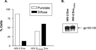
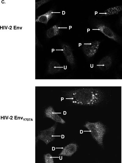
The envelope (Env) protein of human immunodeficiency virus type 2 (HIV-2) and the HIV-1 Vpu protein stimulate the release of retroviral particles from human cells that restrict virus production, an activity that we call the enhancement of virus release (EVR). We have previously shown that two separate domains in the HIV-2 envelope protein are required for this activity: a glycine-tyrosine-x-x-hydrophobic (GYxxtheta) motif in the cytoplasmic tail and an unmapped region in the ectodomain of the protein. We here report that the cellular partner of the GYxxtheta motif is the adaptor protein complex AP-2. The mutation of this motif or the depletion of AP-2 by RNA interference abrogated EVR activity and changed the cellular distribution of the Env from a predominantly punctate pattern to a more diffuse distribution. Since the L domain of equine infectious anemia virus (EIAV) contains a Yxxtheta motif that interacts with AP-2, we used both wild-type and L domain-defective particles of HIV-1 and EIAV to examine whether the HIV-2 Env EVR function was analogous to L domain activity. We observed that the production of all particles was stimulated by HIV-2 Env or Vpu, suggesting that the L domain and EVR activities play independent roles in the release of retroviruses. Interestingly, we found that the cytoplasmic tail of the murine leukemia virus (MLV) Env could functionally substitute for the HIV-2 Env tail, but it did so in a manner that did not require a Yxxtheta motif or AP-2. The cellular distribution of the chimeric HIV-2/MLV Env was significantly less punctate than the wild-type Env, although confocal analysis revealed an overlap in the steady-state locations of the two proteins. Taken together, these data suggest that the essential GYxxtheta motif in the HIV-2 Env tail recruits AP-2 in order to direct Env to a cellular pathway or location that is necessary for its ability to enhance virus release but that an alternate mechanism provided by the MLV Env tail can functionally substitute.


Figures

FIG. 1.
FIG. 1.
Effect of RNA interference against AP-2 on EVR activity. (A) HeLa cells were transfected with an siRNA directed against the 50-kDa μ2 subunit of AP-2 (α-AP-2) or a nonfunctional control siRNA. The cells were then transfected with plasmids expressing HIV-1 Gag-Pol-Rev alone (−) or together with plasmids expressing either HIV-2 Env or Vpu (+). Cell lysates and pelleted virions were analyzed by Western blotting by using anti-CA and anti-Env antibodies. (B) Western blot showing knockdown of the 50-kDa μ2 subunit of AP-2 (α-AP-2) in cell lysates following transfection with an anti-AP-2 siRNA. (C) The p24 (CA)-reacting bands in the Western blots were quantitated, and the ratio of the signal in virions to lysates was obtained. The enhancement of budding was calculated by normalizing all ratios to the Gag-Pol-Rev-only sample in the presence of the control siRNA. The results show that AP-2 depletion increases the baseline level of virus release in the absence of EVRs and prevents HIV-2 Env EVR activity, but it has no effect on the activity of Vpu. The data show the average of three independent experiments. Error bars indicate standard error of the mean.
FIG. 2.
FIG. 2.
Position and sequence dependence of the membrane-proximal GYRPV motif. (A) Sequences of the cytoplasmic domains of the test proteins are shown. (B) Representative Western blot showing the intensity of the p24-containing bands in lysates and virions pelleted from supernatants. (C) Quantitation of EVR activity. The enhancement of budding for each sample was calculated as described in the legend for Fig. 1 and normalized to the wild-type HIV-2 Env value (lane 1). The results show that the GYPRV motif is highly position dependent and requires both the glycine and tyrosine residues for activity. Both of these features are hallmarks of lysosome-targeting signals.
FIG. 3.
FIG. 3.
EVR and L domain activities are independent processes. (A) Virion production from both the wild type (WT) and an L domain mutant (LTAL) of HIV-1 responded equally to stimulation by the HIV-2 Env or Vpu (+). The characteristic appearance of the p25 processing intermediate is seen for the L domain mutant, and this defect is not rescued by EVR expression. The enhancement of budding was calculated as described in the legend for Fig. 1, with the Gag-Pol-Rev-only control (−) in each case being normalized to 1 (n = 3). (B) Similar analysis for wild type and L domain mutant (SRSA) of EIAV using anti-p26 (CA) antibody. Error bars indicate standard error of the mean.
FIG.4.
FIG.4.
The Yxxθ motif and AP-2 are important for the cellular distribution of the HIV-2 Env. (A) Relative amounts of two patterns of HIV-2 Env distribution, punctate and diffuse, that were observed in HeLa cells 48 h after transfection of expression plasmids for wild-type HIV-2 Env or the Y707A mutant. Sixty-five cells were scored blind in each arm of the experiment. (B) Western blot analysis demonstrates equivalent levels of expression of the wild type and Y707A mutant of HIV-2 Env in HeLa cell lysates. (C) Representative fields of cells expressing either wild-type Env or the Y707A mutant, showing punctate and diffuse staining patterns. The spots for the Y707A mutant were noticeably larger than those observed with the wild-type Env. Nuclei were stained with DAPI. D, diffuse; P, punctate; U, cells not expressing Env. (D) Effect of knockdown of AP-2 by RNA interference on staining pattern of HIV-2 Env. (E) Representative cells transfected with HIV-2 Env and control and anti-AP-2 siRNAs and stained with anti-Env antibody.
FIG.4.
FIG.4.
The Yxxθ motif and AP-2 are important for the cellular distribution of the HIV-2 Env. (A) Relative amounts of two patterns of HIV-2 Env distribution, punctate and diffuse, that were observed in HeLa cells 48 h after transfection of expression plasmids for wild-type HIV-2 Env or the Y707A mutant. Sixty-five cells were scored blind in each arm of the experiment. (B) Western blot analysis demonstrates equivalent levels of expression of the wild type and Y707A mutant of HIV-2 Env in HeLa cell lysates. (C) Representative fields of cells expressing either wild-type Env or the Y707A mutant, showing punctate and diffuse staining patterns. The spots for the Y707A mutant were noticeably larger than those observed with the wild-type Env. Nuclei were stained with DAPI. D, diffuse; P, punctate; U, cells not expressing Env. (D) Effect of knockdown of AP-2 by RNA interference on staining pattern of HIV-2 Env. (E) Representative cells transfected with HIV-2 Env and control and anti-AP-2 siRNAs and stained with anti-Env antibody.
FIG. 5.
FIG. 5.
(A) Amino acid sequence of the Moloney MLV Env cytoplasmic tail, showing the extent of the R peptide and the location of a Yxxθ motif centered around tyrosine-622, which was mutated to alanine in the E2M2T(MLV*) chimera. (B) Lysates and virus supernatants probed with anti-CA antibody revealed enhancement of HIV-1 budding induced by the HIV-2 Env, the chimera E2M2T(MLV) containing the MLV tail, and chimera E2M2T(MLV*), which contains a Y622A substitution in the cytoplasmic tail. One representative Western blot is shown, together with a graph showing the average of two independent experiments. The anti-p24 (CA) antibody used also reacts with unprocessed p55Gag and processing intermediates such as p41 (MA-CA). Error bars indicate standard error of the mean. (C) Western blot analysis of cell lysates and virus released from HeLa cells that were transfected with control or anti-AP-2 siRNAs. For the graphical representation, enhancement was calculated as described in the legend for Fig. 1, with all values normalized to the Gag-Pol-Rev-only (−) sample receiving the control siRNA.

Similar articles

Cited by

References

    1. Abada, P., B. Noble, and P. M. Cannon. 2005. Functional domains within the human immunodeficiency virus type 2 envelope protein required to enhance virus production. J. Virol. 79:3627-3638. - PMC - PubMed
    1. Balliet, J. W., D. L. Kolson, G. Eiger, F. M. Kim, K. A. McGann, A. Srinivasan, and R. Collman. 1994. Distinct effects in primary macrophages and lymphocytes of the human immunodeficiency virus type 1 accessory genes vpr, vpu, and nef: mutational analysis of a primary HIV-1 isolate. Virology 200:623-631. - PubMed
    1. Batonick, M., M. Favre, M. Boge, P. Spearman, S. Honing, and M. Thali. 2005. Interaction of HIV-1 Gag with the clathrin-associated adaptor AP-2. Virology 342:190-200. - PubMed
    1. Boge, M., S. Wyss, J. S. Bonifacino, and M. Thali. 1998. A membrane-proximal tyrosine-based signal mediates internalization of the HIV-1 envelope glycoprotein via interaction with the AP-2 clathrin adaptor. J. Biol. Chem. 273:15773-15778. - PubMed
    1. Bonifacino, J. S., and L. M. Traub. 2003. Signals for sorting of transmembrane proteins to endosomes and lysosomes. Annu. Rev. Biochem. 72:395-447. - PubMed

Publication types

MeSH terms