. 2006 Mar;80(6):2933-40.
doi: 10.1128/JVI.80.6.2933-2940.2006.

Rescue of infectious rift valley fever virus entirely from cDNA, analysis of virus lacking the NSs gene, and expression of a foreign gene

Affiliations

Rescue of infectious rift valley fever virus entirely from cDNA, analysis of virus lacking the NSs gene, and expression of a foreign gene

Tetsuro Ikegami et al. J Virol. 2006 Mar.

Abstract

Rift Valley fever virus (RVFV) (genus Phlebovirus, family Bunyaviridae) has a tripartite negative-strand genome, causes a mosquito-borne disease that is endemic in sub-Saharan African countries and that also causes large epidemics among humans and livestock. Furthermore, it is a bioterrorist threat and poses a risk for introduction to other areas. In spite of its danger, neither veterinary nor human vaccines are available. We established a T7 RNA polymerase-driven reverse genetics system to rescue infectious clones of RVFV MP-12 strain entirely from cDNA, the first for any phlebovirus. Expression of viral structural proteins from the protein expression plasmids was not required for virus rescue, whereas NSs protein expression abolished virus rescue. Mutants of MP-12 partially or completely lacking the NSs open reading frame were viable. These NSs deletion mutants replicated efficiently in Vero and 293 cells, but not in MRC-5 cells. In the latter cell line, accumulation of beta interferon mRNA occurred after infection by these NSs deletion mutants, but not after infection by MP-12. The NSs deletion mutants formed larger plaques than MP-12 did in Vero E6 cells and failed to shut off host protein synthesis in Vero cells. An MP-12 mutant carrying a luciferase gene in place of the NSs gene replicated as efficiently as MP-12 did, produced enzymatically active luciferase during replication, and stably retained the luciferase gene after 10 virus passages, representing the first demonstration of foreign gene expression in any bunyavirus. This reverse genetics system can be used to study the molecular virology of RVFV, assess current vaccine candidates, produce new vaccines, and incorporate marker genes into animal vaccines.


Figures

FIG. 1.
FIG. 1.
Introduction of gene marker into S, M, and L segments. (A) Alignment of nucleotide and amino acid (aa.) sequences. Introduced XhoI sites on virus-sense S (Sv), anti-virus-sense M (Mvc), and anti-virus-sense L (Lvc) segments were underlined. Mutation positions were shown as arrowheads. (B) Demonstration of the XhoI marker in rMP-12 RNA. Viral RNA was extracted from culture supernatants of Vero E6 cells infected with MP-12 and rMP-12 in the experiment listed in Table 1, plasmid combination A, experiment 1 (A: Exp.1). After the RNA was digested with DNase I at 37°C for 1 h, PCR was performed with (+) and without (−) the reverse transcription step (RT). Water was used as a negative control. Digestion with XhoI was performed at 37°C for 2 h. The expected sizes of XhoI-digested fragments are shown to the right of the gels. The positions of molecular size markers are shown to the left of the gels. (C) Demonstration of XhoI marker in rMP-12 recovered without using protein expression plasmids in the experiment listed in Table 1, plasmid combination D, experiment 1 (D: Exp.1).
FIG. 2.
FIG. 2.
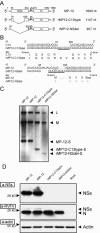
Production of NSs deletion mutants. (A) Diagrams of NSs deletion mutants. (B) Alignment of nucleotide (nt.) and amino acid (aa.) sequences of clone 13 (C13) and rMP12-C13type or of MP-12 and rMP12-NSdel. Additional sequences in the mutants are underlined. The rMP12-NSdel does not contain the first AUG (methionine [M]) and does not produce NSs protein. N-term, N-terminal; C-term, C-terminal. (C) Purified virion RNAs of MP-12, rMP-12, rMP12-C13type, and rMP12-NSdel were analyzed by Northern blotting using virus-sense S-, M-, and L-specific RNA probes (8). (D) Western blotting using anti-NSs (a-NSs), anti-RVFV (a-RVFV), and anti-actin (a-actin) antibodies (8). Vero E6 cells were infected with MP-12 and mutants at an MOI of 1 and were harvested at 6 h p.i.
FIG. 3.
FIG. 3.
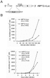
Production of recombinant MP-12 expressing Renilla luciferase. (A) Diagram of the S segment of rMP12-rLuc. Renilla luciferase ORF with HpaI and SpeI sites at 5′ and 3′ ends, respectively, was inserted into the cassette of S segment digested with HpaI and SpeI. (B) Vero E6 cells were infected with rMP12-rLuc or MP-12 at the indicated MOI, and luciferase (Luc.) activity was tested at several time points as shown. Luciferase activities of 3.2 × 10⁴ cells and 1.6 × 10³ cells are shown for early (top panel) and late (bottom panel) time points, respectively.
FIG. 4.
FIG. 4.
Growth curves of MP-12 and mutants. Vero cells (A), 293 cells (B), and MRC-5 cells (C and D) were infected with MP-12, rMP-12, rMP12-C13type, rMP12-NSdel, and rMP12-rLuc at an MOI of 1 (A, B, and C) or 0.01 (D), and the culture supernatants were collected at the time points shown. Culture supernatants were collected from three independent wells at each time point, and the virus titers were determined in Vero E6 cells by assay. The values are mean titers ± standard deviations (error bars) from three independent experiments.
FIG. 5.
FIG. 5.
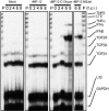
Accumulation of IFN-β mRNA and TNF-α mRNA in MRC-5 cells infected with NSs deletion mutants. MRC-5 cells were mock infected (Mock) or infected with rMP-12, rMP12-C13type, and rMP12-NSdel at an MOI of 1. Total intracellular RNAs were extracted at the indicated times (hours p.i. [h.p.i.]). RNA samples were hybridized with multiprobe template (hCK-3), and RNase protection assay was performed. P, probes; TGFβ3, transforming growth factor β3; GAPDH, glyceraldehyde-3-phosphate dehydrogenase.
FIG. 6.
FIG. 6.
Responses of host cells infected with MP-12 and mutants. (A) Plaque production in Vero E6 cells by MP-12, rMP-12, rMP12-C13type, and rMP12-NSdel stained with neutral red and crystal violet. (B) Shutoff of host protein synthesis. Vero cells were mock infected (Mock) or infected with MP-12, rMP-12, rMP12-C13type, and rMP12-NSdel at an MOI of 5. Cells were labeled with 100 μCi/ml of [35S]methionine for 1 h at 17 h postinfection. Cell extracts were analyzed on 10% polyacrylamide gel. The positions of synthesized N and NSs proteins are shown by arrowheads to the right of the gel.

References

    1. Billecocq, A., M. Spiegel, P. Vialat, A. Kohl, F. Weber, M. Bouloy, and O. Haller. 2004. NSs protein of Rift Valley fever virus blocks interferon production by inhibiting host gene transcription. J. Virol. 78:9798-9806. - PMC - PubMed
    1. Blakqori, G., and F. Weber. 2005. Efficient cDNA-based rescue of La Crosse bunyaviruses expressing or lacking the nonstructural protein NSs. J. Virol. 79:10420-10428. - PMC - PubMed
    1. Bouloy, M., C. Janzen, P. Vialat, H. Khun, J. Pavlovic, M. Huerre, and O. Haller. 2001. Genetic evidence for an interferon-antagonistic function of Rift Valley fever virus nonstructural protein NSs. J. Virol. 75:1371-1377. - PMC - PubMed
    1. Bridgen, A., and R. M. Elliott. 1996. Rescue of a segmented negative-strand RNA virus entirely from cloned complementary DNAs. Proc. Natl. Acad. Sci. USA 93:15400-15404. - PMC - PubMed
    1. Bridgen, A., F. Weber, J. K. Fazakerley, and R. M. Elliott. 2001. Bunyamwera bunyavirus nonstructural protein NSs is a nonessential gene product that contributes to viral pathogenesis. Proc. Natl. Acad. Sci. USA 98:664-669. - PMC - PubMed

Publication types

MeSH terms

LinkOut - more resources