. 2006 Mar;80(6):2949-57.
doi: 10.1128/JVI.80.6.2949-2957.2006.

Infection of dendritic cells (DCs), not DC-SIGN-mediated internalization of human immunodeficiency virus, is required for long-term transfer of virus to T cells

Affiliations

Infection of dendritic cells (DCs), not DC-SIGN-mediated internalization of human immunodeficiency virus, is required for long-term transfer of virus to T cells

Laura Burleigh et al. J Virol. 2006 Mar.

Abstract

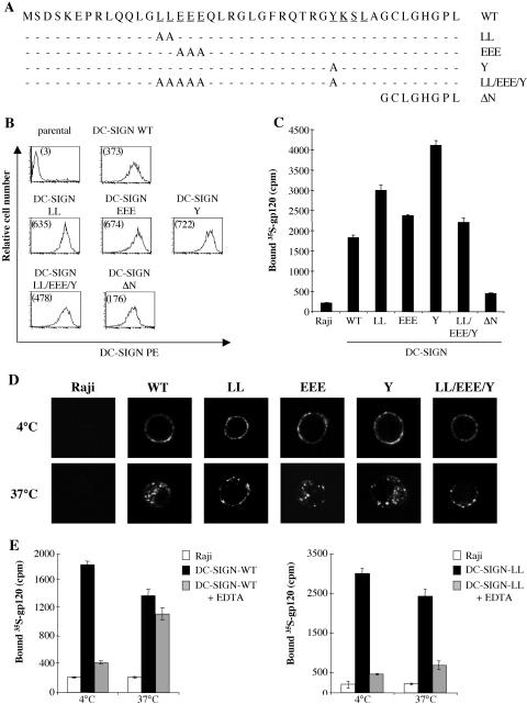
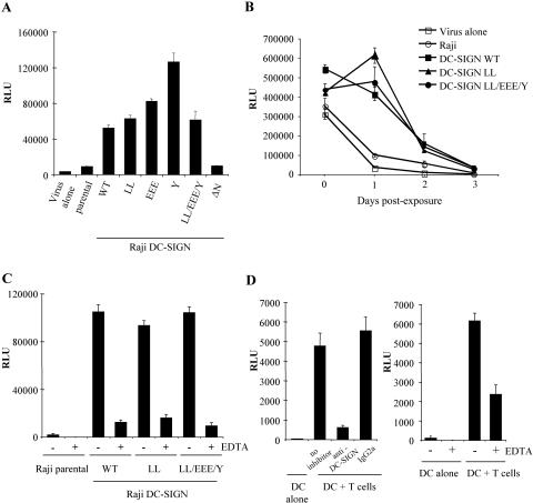
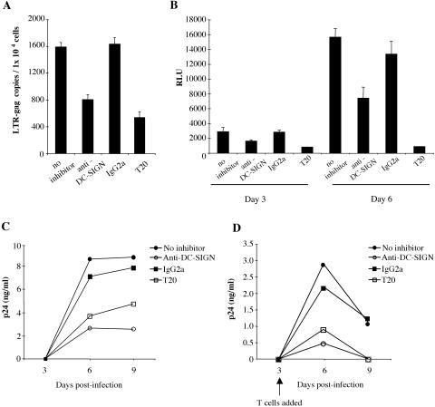
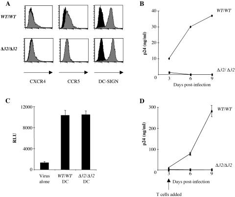
The C-type lectin DC-SIGN expressed on immature dendritic cells (DCs) captures human immunodeficiency virus (HIV) particles and enhances the infection of CD4+ T cells. This process, known as trans-enhancement of T-cell infection, has been related to HIV endocytosis. It has been proposed that DC-SIGN targets HIV to a nondegradative compartment within DCs and DC-SIGN-expressing cells, allowing incoming virus to persist for several days before infecting target cells. In this study, we provide several lines of evidence suggesting that intracellular storage of intact virions does not contribute to HIV transmission. We show that endocytosis-defective DC-SIGN molecules enhance T-cell infection as efficiently as their wild-type counterparts, indicating that DC-SIGN-mediated HIV internalization is dispensable for trans-enhancement. Furthermore, using immature DCs that are genetically resistant to infection, we demonstrate that several days after viral uptake, HIV transfer from DCs to T cells requires viral fusion and occurs exclusively through DC infection and transmission of newly synthesized viral particles. Importantly, our results suggest that DC-SIGN participates in this process by cooperating with the HIV entry receptors to facilitate cis-infection of immature DCs and subsequent viral transfer to T cells. We suggest that such a mechanism, rather than intracellular storage of incoming virus, accounts for the long-term transfer of HIV to CD4+ T cells and may contribute to the spread of infection by DCs.


Figures

FIG. 1.
FIG. 1.
Characterization of WT and mutant DC-SIGN proteins. (A) Schematic representation of mutations introduced into the DC-SIGN cytoplasmic tail. The dileucine (LL) motif, tyrosine-based (YSKL) motif, and triacidic cluster (EEE) are underlined. (B) FACS analysis of Raji cells expressing either WT or mutated forms of DC-SIGN. The mean fluorescence intensities are indicated in parentheses. (C) HIV gp120 binding. [35S]gp120 (25 nM) was incubated for 1 h at 4°C with Raji and Raji-DC-SIGN cells. Cells were washed four times before measurements of cell-associated radioactivity. (D) Subcellular localization of WT and mutant DC-SIGN. Anti-DC-SIGN MAb 8A5 was incubated with Raji or Raji-DC-SIGN WT cells or LL, Y, or EEE mutant cells for 1 h at 4°C. Cells were washed to eliminate unbound antibody and incubated for 30 min at either 4°C or 37°C. DC-SIGN-bound 8A5 was detected with a fluorescein isothiocyanate-conjugated secondary Ab. (E) DC-SIGN-mediated gp120 internalization. 35S-labeled gp120 was bound to Raji, Raji-DC-SIGN WT (left panel), or Raji-DC-SIGN LL mutant (right panel) cells for 1 h at 4°C. Cells were extensively washed and incubated for 30 min at either 4°C or 37°C. Cells were treated with EDTA or mock treated to distinguish internalized from cell surface-bound gp120. Cells were then washed three times before measurements of cell-associated radioactivity.
FIG. 2.
FIG. 2.
DC-SIGN-mediated enhancement of T-cell infection does not require DC-SIGN internalization. (A) Enhancement of T-cell infection. Raji or Raji-DC-SIGN cells were incubated with JR-CSF Luc at a low MOI (0.1) for 2 h at 37°C and cocultured with activated T cells. Viral replication was assessed by measuring luciferase activities in cell lysates at 3 days postinfection. As a control, activated T cells were incubated with virus alone. RLU, relative light units. (B) Retention of viral infectivity. Raji and DC-SIGN-expressing cells were pulsed with JR-CSF (MOI, 0.1) at 37°C, and aliquots of cells were added daily to 293T cells expressing CD4 and CCR5. Luciferase activities were determined 3 days later. (C) Raji and Raji-DC-SIGN cells were incubated with JR-CSF Luc (MOI, 0.1) for 3 h at 37°C. Cells were treated for 10 min with 20 mM EDTA to remove virus bound to DC-SIGN at the cell surface or were mock treated with PBS. After being washed extensively, cells were cocultured with 293T cells expressing CD4 and CCR5, and luciferase activities were determined 3 days later. (D) (Left panel) Immature DCs were pulsed with HIV Luc pseudotyped with the HXB2 envelope (10 ng of p24) for 2 hours at 37°C. Cells were treated with 20 mM EDTA for 10 min or mock treated with PBS, washed extensively, and cocultured with activated T cells for 3 days before determining the luciferase activity. (Right panel) DCs were pretreated with anti-DC-SIGN MAb (20 μg/ml) or an isotype control (20 μg/ml) for 20 min, washed extensively, and pulsed with HIV Luc pseudotyped with the HXB2 envelope (10 ng of p24) for 2 hours at 37°C. Cells were washed and cocultured with activated T cells for 3 days before determining the luciferase activity. Data are representative of three independent experiments, and values are given as means of triplicates ± standard deviations.
FIG. 3.
FIG. 3.
DC-SIGN-mediated cis-infection of DCs contributes to long-term transfer of HIV to CD4+ T cells. (A) HIV DNA synthesis. DCs (1 × 106) were preincubated with either anti-DC-SIGN MAb (25 μg/ml), an isotype control (25 μg/ml), or T20 (1 μM) or were mock treated. Cells were then infected with the HIV X4 strain NL43 (100 ng of p24) for 4 hours at 37°C, washed extensively, and maintained in culture for 2 days. Cells were harvested, and late viral DNA products were quantified by real-time PCR. (B) DC infection by X4 HIV Luc. Immature DCs were pretreated with inhibitors as described for panel A. Cells were then infected with single-cycle HIV Luc pseudotyped with the HXB2 envelope (10 ng of p24) for 3 h at 37°C. DCs were washed extensively and maintained in RPMI supplemented with GM-CSF and IL-4 for either 3 or 6 days before determining the luciferase activity. (C) DCs were pretreated with inhibitors as described for panel A before the addition of NLAD8 (0.4 ng of p24 for 1 × 10⁵ cells) for 3 h at 37°C. Cells were washed extensively and maintained in culture. p24 Gag accumulation in cell supernatants was determined on days 3, 6, and 9. (D) DCs were infected as described for panel C. Three days after infection, DCs were cocultured with 1 × 10⁵ activated primary T cells. Aliquots of culture supernatants were taken at the indicated time points and assayed for p24. The experiments presented here were carried out using DCs generated from different donors. Data are representative of three independent experiments, and values are given as means of triplicates ± standard deviations.
FIG. 4.
FIG. 4.
DC infection is essential for delayed transfer of HIV to T cells. (A) FACS analysis of cell surface expression of CXCR4, CCR5, and DC-SIGN on WT/WT and Δ32/Δ32 immature DCs. Black histograms show data for an isotype control Ab. (B) DC infection. WT/WT and Δ32/Δ32 DCs (4 × 10⁵) were infected with the R5 strain NLAD8 (10 ng of p24) for 3 h at 37°C. Cells were washed and cultured in medium containing GM-CSF and IL-4. Viral p24 Gag in the culture supernatants was measured on days 3, 6, and 9 postinfection. (C) Short-term transfer. WT/WT and Δ32/Δ32 immature DCs were infected at a low MOI (0.1) with R5 BaL Luc pseudotyped viruses for 2 hours and cocultured with activated T cells. Luciferase activities in cell lysates were evaluated at 3 days postinfection. As a control, HIV Luc viruses were incubated with medium alone (virus alone) and, 2 hours later, transferred to T cells. (D) Long-term transfer. WT/WT and Δ32/Δ32 DCs (4 × 10⁵) were infected with the R5 HIV strain NLAD8 (10 ng of p24) for 3 h at 37°C. Cells were washed and cultured in RPMI-GM-CSF-IL-4 for 3 days before the addition of an equal number of activated T cells. Aliquots of culture supernatants were taken at the indicated time points and assayed for p24 antigen by enzyme-linked immunosorbent assay. Data are representative of two independent experiments using two different Δ32/Δ32 donors.

Similar articles

Cited by

References

    1. Amara, A., A. Vidy, G. Boulla, K. Mollier, J. Garcia-Perez, J. Alcami, C. Blanpain, M. Parmentier, J. L. Virelizier, P. Charneau, and F. Arenzana-Seisdedos. 2003. G protein-dependent CCR5 signaling is not required for efficient infection of primary T lymphocytes and macrophages by R5 human immunodeficiency virus type 1 isolates. J. Virol. 77:2550-2558. - PMC - PubMed
    1. Canque, B., Y. Bakri, S. Camus, M. Yagello, A. Benjouad, and J. C. Gluckman. 1999. The susceptibility to X4 and R5 human immunodeficiency virus-1 strains of dendritic cells derived in vitro from CD34(+) hematopoietic progenitor cells is primarily determined by their maturation stage. Blood 93:3866-3875. - PubMed
    1. Curtis, B. M., S. Scharnowske, and A. J. Watson. 1992. Sequence and expression of a membrane-associated C-type lectin that exhibits CD4-independent binding of human immunodeficiency virus envelope glycoprotein gp120. Proc. Natl. Acad. Sci. USA 89:8356-8360. - PMC - PubMed
    1. Deng, H. K., D. Unutmaz, V. N. KewalRamani, and D. R. Littman. 1997. Expression cloning of new receptors used by simian and human immunodeficiency viruses. Nature 388:296-300. - PubMed
    1. Engering, A., T. B. Geijtenbeek, S. J. van Vliet, M. Wijers, E. van Liempt, N. Demaurex, A. Lanzavecchia, J. Fransen, C. G. Figdor, V. Piguet, and Y. van Kooyk. 2002. The dendritic cell-specific adhesion receptor DC-SIGN internalizes antigen for presentation to T cells. J. Immunol. 168:2118-2126. - PubMed

Publication types

MeSH terms

Substances