. 2006 Mar;80(6):3050-61.
doi: 10.1128/JVI.80.6.3050-3061.2006.

A hydrophobic binding surface on the human immunodeficiency virus type 1 Nef core is critical for association with p21-activated kinase 2

Affiliations

A hydrophobic binding surface on the human immunodeficiency virus type 1 Nef core is critical for association with p21-activated kinase 2

Kristin Agopian et al. J Virol. 2006 Mar.

Abstract

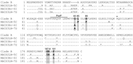
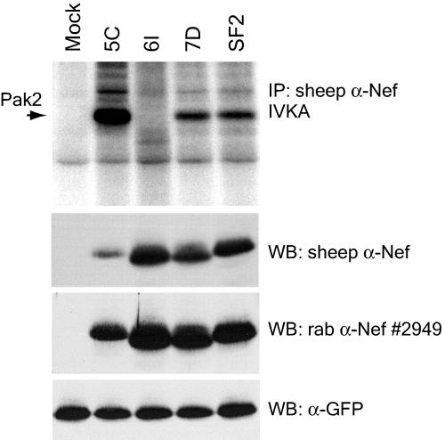
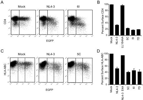
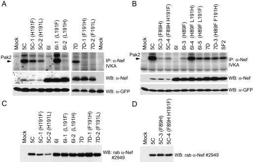
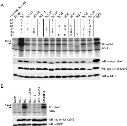
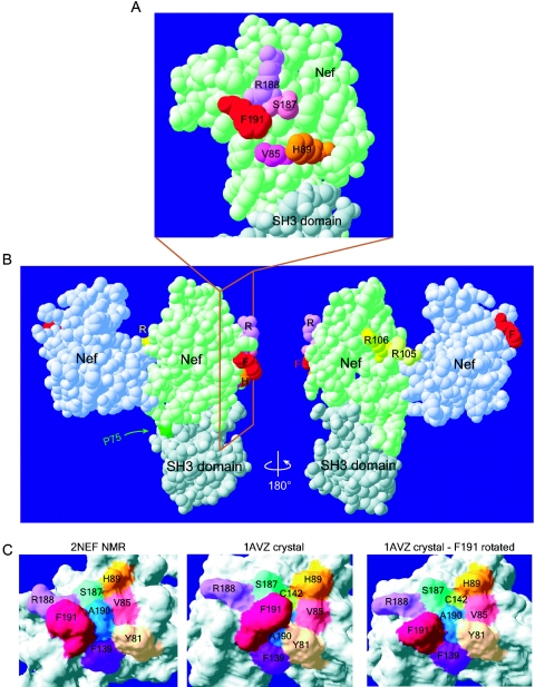
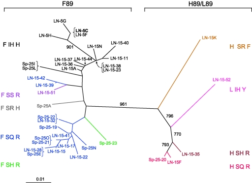
The interaction of human immunodeficiency virus type 1 (HIV-1) Nef with p21-activated kinase 2 (Pak2) has been proposed to play an important role in T-cell activation and disease progression during viral infection. However, the mechanism by which Nef activates Pak2 is poorly understood. Mutations in most Nef motifs previously reported to be required for Pak2 activation (G2, PxxP72, and RR105) also affect other Nef functions, such as CD4 or major histocompatibility complex class I (MHC-I) downregulation. To better understand Nef interactions with Pak2, we performed mutational analysis of three primary HIV-1 Nef clones that exhibited similar capacities for downregulation of CD4 and MHC-I but variable abilities to associate with activated Pak2. Our results demonstrate that Nef amino acids at positions 85, 89, 187, 188, and 191 (L, H, S, R, and F in the clade B consensus, respectively) are critical for Pak2 association. Mutation of these Nef residues dramatically altered association with Pak2 without affecting Nef expression levels or CD4 and MHC-I downregulation. Furthermore, compensation occurred at positions 89 and 191 when both amino acids were substituted. Since residues 85, 89, 187, 188, and 191 cluster on the surface of the Nef core domain in a region distinct from the dimerization and SH3-binding domains, we propose that these Nef residues form part of a unique binding surface specifically involved in association with Pak2. This binding surface includes exposed and recessed hydrophobic residues and may participate in an as-yet-unidentified protein-protein interaction to facilitate Pak2 activation.


Figures

FIG. 1.
FIG. 1.
Amino acid alignment of primary HIV-1 Nef clones compared to the clade B consensus. Amino acids G83, L85, H89, S187, R188, and F191, numbered relative to the NL4-3 Nef sequence, are shaded. Boldface type indicates positions that vary from the consensus. Also labeled are R106, previously proposed to be important for Pak2 activation (51, 54), and the PxxP SH3-binding domain. The clade B consensus is from the LANL sequence database (August 2004). Alanine is the consensus amino acid at position 83 of an earlier LANL clade B Nef consensus sequence.
FIG. 2.
FIG. 2.
Associations of primary HIV-1 Nefs with activated Pak2 are variable. 293T cells transiently expressing GFP and the indicated Nefs were immunoprecipitated with sheep anti-Nef (α-Nef) (raised to SF2 Nef) and assayed by IVKA (top panel) as described in Materials and Methods. SF2 Nef, previously demonstrated to strongly activate Pak2 (4), is included as a positive control. An arrow indicates the 62-kDa band corresponding to Pak2. Whole-cell lysates used for IVKA immunoprecipitations (IP) were immunoblotted with sheep anti-Nef (second panel) or rabbit (rab) anti-Nef antibody no. 2949. As a control for transfection efficiency, lysates were immunoblotted for GFP expression (bottom panel). WB, Western blot.
FIG. 3.
FIG. 3.
Downregulation of cell surface CD4 and MHC-I by primary Nef alleles is conserved. Flow cytometric analysis of CD4 downregulation (A and B) and MHC-I downregulation (C and D). (A) Flow cytometric analysis of HIJ cells cotransfected with pCDNA3-EGFP and either pCR3.1 (Mock), pCR3.1-Nef-NL4-3 (NL4-3), or pCR3.1-Nef-MACS3Br-6I (6I). Vertical lines indicate the threshold for GFP expression. Data shown are representative of two independent experiments. (B) Quantitation of CD4 downregulation data. Percent surface CD4 was calculated from the geometric mean fluorescence in GFP-positive cells as described in Materials and Methods and is shown relative to that for GFP-positive mock-transfected cells. The LL164AA mutation, previously shown to abolish CD4 downregulation, was made in the background of NL4-3 Nef and was included as a negative control. Averages are from two independent experiments. Error bars represent standard deviations. (C) Flow cytometric analysis of Jurkat T-antigen cells cotransfected with EGFP and either pCR3.1 (Mock), NL4-3 Nef, or 5C Nef. Surface MHC-I levels were determined by staining with anti-HLA-ABC-PE. Vertical lines indicate gates for GFP-negative, low-GFP-expressing, and high-GFP-expressing cells. Data are representative of four independent experiments. (D) Quantitation of MHC-I downregulation. Percent surface MHC-I was calculated as described above but includes only the high-GFP-expressing cells (right panels in dot plots). E4A is the EEEE65AAAA mutation of NL4-3 Nef, a mutation that has been shown to abolish MHC-I downregulation (46) and was included as a negative control. Averages are from one to four independent experiments. Error bars represent standard deviations.
FIG. 4.
FIG. 4.
Amino acids 89 and 191 of Nef are critical for Pak2 association, and mutations at these positions are compensatory in the 6I and 7D Nef backgrounds. (A and B) IVKAs. Whole-cell lysates of 293T cells transiently expressing GFP and single and double mutants of 5C, 6I, and 7D Nefs were immunoprecipitated with sheep anti-Nef (α-Nef) and assayed by IVKA (top panel) as described in Materials and Methods. An arrow indicates the 62-kDa band corresponding to Pak2. Whole-cell lysates used for IVKA immunoprecipitations (IP) were immunoblotted with rabbit (rab) or sheep anti-Nef antibodies (raised to SF2) (middle panel) or anti-GFP (bottom panel). (C and D) Independent transfection experiments to confirm relative expression levels of Nef mutants. 293T cells were transfected with GFP and Nef as described for panels A and B. Whole-cell lysates were immunoblotted with rabbit anti-Nef antibody no. 2949. WB, Western blot.
FIG. 5.
FIG. 5.
Amino acids F85, I187, and H188 are specifically required for association of 5C Nef with Pak2. (A and B) IVKAs. Whole-cell lysates of 293T cells transiently expressing GFP and expressing 5C Nef mutants were immunoprecipitated with sheep anti-Nef and assayed by IVKA (top panels) as described in Materials and Methods. An arrow indicates the 62-kDa band corresponding to Pak2. Whole-cell lysates used for IVKA immunoprecipitations (IP) were immunoblotted with sheep anti-Nef (α-Nef) (raised to SF2), rabbit (rab) anti-Nef antibody no. 2949 (middle panels), or anti-GFP (bottom panels). The chart at the top of panel A indicates the amino acid changes made for each mutant. All lanes shown in panel B are from the same experiment. WB, Western blot.
FIG. 6.
FIG. 6.
Mutation F89H abolishes association of 5C Nef with exogenous Pak2. 293T cells were cotransfected with FLAG-tagged, kinase-dead Pak2-K278R, dominant active Cdc42-V12, and either vector alone (Mock), 5C, or mutant 5C-3. Cell lysates were immunoprecipitated with sheep anti-Nef (α-Nef) antibody. Nef-associated FLAG-Pak2-K278R was detected by anti-FLAG antibody (top panel), and immunoprecipitated Nef was detected by rabbit (rab) anti-Nef antibody no. 2949 (second panel). Expression of FLAG-Pak2-K278R and Nef in cell lysates was assayed by anti-FLAG and anti-Nef Western blotting (third and fourth panels). Results are representative of two independent experiments. IP, immunoprecipitation; WB, Western blot.
FIG. 7.
FIG. 7.
Residues 85, 89, 187, 188, and 191 cluster together on the surface of the Nef core domain in a region distinct from the dimerization and SH3-binding domains. Computer modeling of Nef core domain structures demonstrates the proximity of amino acids important for Pak2 association. Structures were obtained from the Protein Data Bank and were visualized with Swiss PDB viewer DeepView. (A and B) van der Waals surfaces of the 1AVZ Nef crystal structure (2). The Nef core domain (light green) is bound to a second Nef core (light blue) and to the Fyn kinase SH3 domain (gray). The Nef core domain comprises amino acids 71 to 148 and 179 to 203. (A) Residues 85 (fuchsia), 89 (orange), 187 (pink), 188 (violet), and 191 (red) cluster together on the surface of the Nef core domain, with phenylalanine 191 protruding from the surface. The SH3 domain of Fyn is shown binding to the PxxP domain of Nef. Panel A is an enlargement, rotated 90°, of the boxed region in panel B. (B) F191 (red) is distant from the dimerization surface and arginines 105 and 106 (yellow). P75 (dark green) is part of the PxxP SH3-binding domain. (C) F191 of Nef can adopt two different confomers that either expose or hide the recessed A190 (dark blue). (Left) The molecular surface of the 2NEF NMR model of the Nef core domain was computed with DeepView (22). The confomer adopted by F191 in this model occurs in 50% of the 40 published NMR models, while the other 50% are similar to that shown in the middle panel. (Middle) Molecular surface of the Nef core domain of the 1AVZ crystal structure (2). Here, contact with a neighboring molecule in the crystal lattice sterically hinders F191 and constrains it into adopting the confomer shown. (Right) Molecular surface of the 1AVZ crystal structure with the side chain of F191 rotated to adopt a confomer, as in 2NEF. No steric hindrances occurred with this confomer.
FIG. 8.
FIG. 8.
Phylogenetic analysis of 37 full-length Nef clones from patient MACS2 reveals distinct populations that segregate according to variation at residue 89. Full-length Nef amino acid sequences amplified by PCR from the MACS2LN viral isolate (4 sequences) or directly from autopsy lymph node (22 sequences) or spleen tissue (11 sequences) were aligned with Clustal W. Clones are colored according to their amino acid sequence at residues 89, 187, 188, and 191, as indicated to the left and right of the tree. Clustal X was used to create the bootstrapped, neighbor-joining tree. The numbers associated with each branch represent the bootstrap values, which indicate the reliability of the branching order and were derived from 1,000 replicates.

References

    1. Alexander, L., Z. Du, M. Rosenzweig, J. U. Jung, and R. C. Desrosiers. 1997. A role for natural simian immunodeficiency virus and human immunodeficiency virus type 1 Nef alleles in lymphocyte activation. J. Virol. 71:6094-6099. - PMC - PubMed
    1. Arold, S., P. Franken, M. P. Strub, F. Hoh, S. Benichou, R. Benarous, and C. Dumas. 1997. The crystal structure of HIV-1 Nef protein bound to the Fyn kinase SH3 domain suggests a role for this complex in altered T cell receptor signaling. Structure 5:1361-1372. - PubMed
    1. Arold, S. T., and A. S. Baur. 2001. Dynamic Nef and Nef dynamics: how structure could explain the complex activities of this small HIV protein. Trends Biochem. Sci. 26:356-363. - PubMed
    1. Arora, V. K., R. P. Molina, J. L. Foster, J. L. Blakemore, J. Chernoff, B. L. Fredericksen, and J. V. Garcia. 2000. Lentivirus Nef specifically activates Pak2. J. Virol. 74:11081-11087. - PMC - PubMed
    1. Bagrodia, S., and R. A. Cerione. 1999. Pak to the future. Trends Cell Biol. 9:350-355. - PubMed

Publication types

MeSH terms

Substances

Associated data