

Perforin expression in the gastrointestinal mucosa is limited to acute simian immunodeficiency virus infection
- PMID: 16501118
- PMCID: PMC1395471
- DOI: 10.1128/JVI.80.6.3083-3087.2006
Perforin expression in the gastrointestinal mucosa is limited to acute simian immunodeficiency virus infection
Abstract
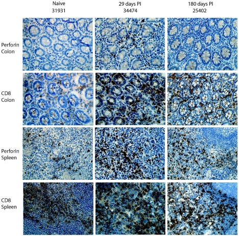
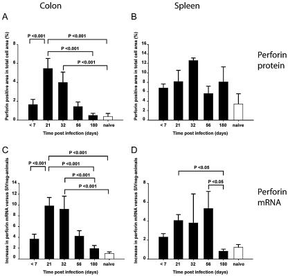
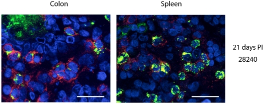
Perforin-mediated cytotoxicity is a major effector function of virus-specific CD8 T cells. We have investigated the expression of perforin in the gut, an important site of simian immunodeficiency virus (SIV) pathogenesis, during experimental SIV infection of rhesus macaques. We observed significant increases in perforin protein and mRNA expression levels in the colons of SIV-infected macaques as early as 21 days after infection. However, during chronic infection, despite ongoing viral replication, perforin expression returned to levels similar to those detected in SIV-naïve animals. These findings demonstrate the presence of a robust perforin-positive response in gastrointestinal CD8 T cells during acute, but not chronic, SIV infection.
Figures
Similar articles
-
Expansion of FOXP3+ CD8 T cells with suppressive potential in colorectal mucosa following a pathogenic simian immunodeficiency virus infection correlates with diminished antiviral T cell response and viral control.J Immunol. 2010 Feb 15;184(4):1690-701. doi: 10.4049/jimmunol.0902955. Epub 2010 Jan 6. J Immunol. 2010. PMID: 20053943
-
Simian Immunodeficiency Virus-Producing Cells in Follicles Are Partially Suppressed by CD8+ Cells In Vivo.J Virol. 2016 Nov 28;90(24):11168-11180. doi: 10.1128/JVI.01332-16. Print 2016 Dec 15. J Virol. 2016. PMID: 27707919 Free PMC article.
-
Heterogeneity of the simian immunodeficiency virus (SIV) specific CD8(+) T-cell response in mucosal tissues during SIV primary infection.Microbes Infect. 2003 Jul;5(9):757-67. doi: 10.1016/s1286-4579(03)00144-8. Microbes Infect. 2003. PMID: 12850201
-
Cytotoxic T lymphocytes specific for the simian immunodeficiency virus.Immunol Rev. 1999 Aug;170:127-34. doi: 10.1111/j.1600-065x.1999.tb01334.x. Immunol Rev. 1999. PMID: 10566147 Review.
-
The Hitchhiker Guide to CD4+ T-Cell Depletion in Lentiviral Infection. A Critical Review of the Dynamics of the CD4+ T Cells in SIV and HIV Infection.Front Immunol. 2021 Jul 21;12:695674. doi: 10.3389/fimmu.2021.695674. eCollection 2021. Front Immunol. 2021. PMID: 34367156 Free PMC article.
Cited by
-
Tissue issues: mucosal T-cell responses in HIV-1 infection.Curr Opin HIV AIDS. 2019 Mar;14(2):100-107. doi: 10.1097/COH.0000000000000530. Curr Opin HIV AIDS. 2019. PMID: 30601239 Free PMC article. Review.
-
Mechanisms of gastrointestinal CD4+ T-cell depletion during acute and early human immunodeficiency virus type 1 infection.J Virol. 2007 Jan;81(2):599-612. doi: 10.1128/JVI.01739-06. Epub 2006 Oct 25. J Virol. 2007. PMID: 17065209 Free PMC article.
-
HIV Infection and Gut Mucosal Immune Function: Updates on Pathogenesis with Implications for Management and Intervention.Curr Infect Dis Rep. 2010 Jan;12(1):19-27. doi: 10.1007/s11908-009-0072-9. Epub 2010 Jan 13. Curr Infect Dis Rep. 2010. PMID: 20174448 Free PMC article.
-
Defining T Cell Tissue Residency in Humans: Implications for HIV Pathogenesis and Vaccine Design.Curr HIV/AIDS Rep. 2020 Apr;17(2):109-117. doi: 10.1007/s11904-020-00481-7. Curr HIV/AIDS Rep. 2020. PMID: 32052270 Free PMC article. Review.
-
Mucosal immunity in acute HIV: a review of recent work.Curr Opin HIV AIDS. 2025 May 1;20(3):193-198. doi: 10.1097/COH.0000000000000917. Epub 2025 Jan 24. Curr Opin HIV AIDS. 2025. PMID: 39903645 Review.
References
-
- Abel, K., L. La Franco-Scheuch, T. Rourke, Z.-M. Ma, V. de Silva, B. Fallert, L. Beckett, T. A. Reinhart, and C. J. Miller. 2004. Gamma interferon-mediated inflammation is associated with lack of protection from intravaginal simian immunodeficiency virus SIVmac239 challenge in simian-human immunodeficiency virus 89.6-immunized rhesus macaques. J. Virol. 78:841-854. - PMC - PubMed
-
- Bjork, L., T. E. Fehniger, U. Andersson, and J. Andersson. 1996. Computerized assessment of production of multiple human cytokines at the single-cell level using image analysis. J. Leukoc. Biol. 59:287-295. - PubMed
-
- Brenchley, J. M., T. W. Schacker, L. E. Ruff, D. A. Price, J. H. Taylor, G. J. Beilman, P. L. Nguyen, A. Khoruts, M. Larson, A. T. Haase, and D. C. Douek. 2004. CD4+ T cell depletion during all stages of HIV disease occurs predominantly in the gastrointestinal tract. J. Exp. Med. 200:749-759. - PMC - PubMed
-
- Brodie, S. J., D. A. Lewinsohn, B. K. Patterson, D. Jiyamapa, J. Krieger, L. Corey, P. D. Greenberg, and S. R. Riddell. 1999. In vivo migration and function of transferred HIV-1-specific cytotoxic T cells. Nat. Med. 5:34-41. - PubMed
-
- Dailey, P. J., M. Zamround, R. Kelso, J. Kolberg, and M. Urdea. 1995. Quantification of simian immunodeficiency virus (SIV) RNA in plasma of acute and chronically infected macaques using a branched DNA (bDNA) signal amplification assay. Presented at the 13th Annual Symposium on Nonhuman Primate Models for AIDS, Monterey, Calif.
Publication types
MeSH terms
Substances
Grants and funding
LinkOut - more resources
Full Text Sources
Research Materials
