. 2006 Mar;15(3):543-52.
doi: 10.1110/ps.051733506.

Physical characterization of MxiH and PrgI, the needle component of the type III secretion apparatus from Shigella and Salmonella

Affiliations

Physical characterization of MxiH and PrgI, the needle component of the type III secretion apparatus from Shigella and Salmonella

Numukunda Darboe et al. Protein Sci. 2006 Mar.

Abstract

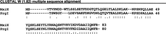
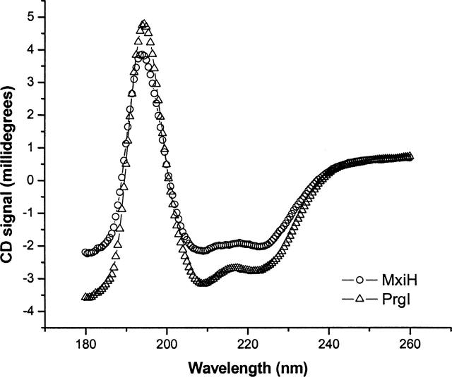
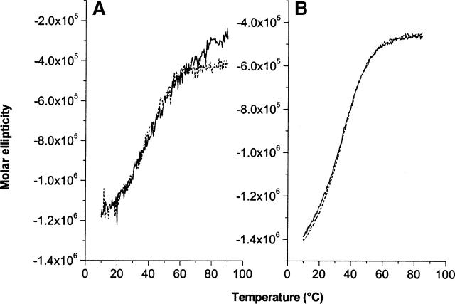
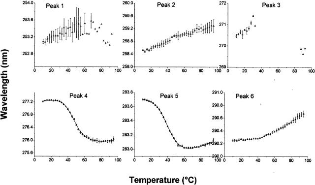
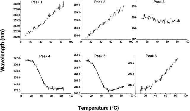
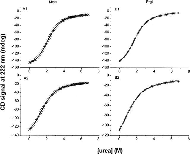
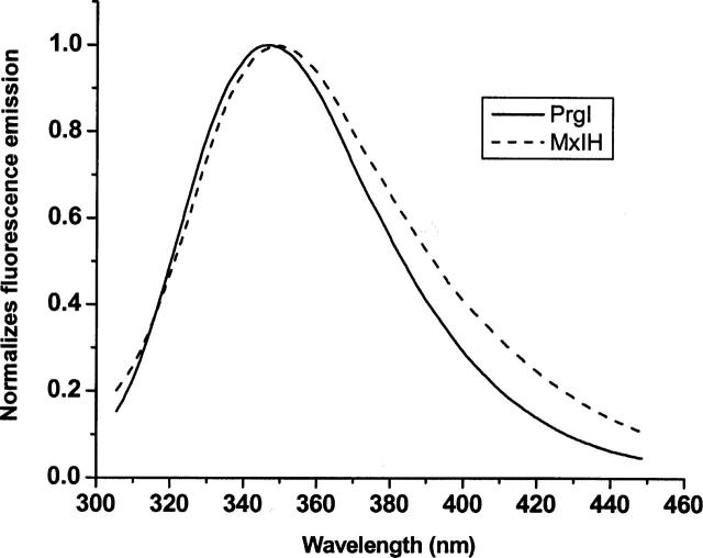
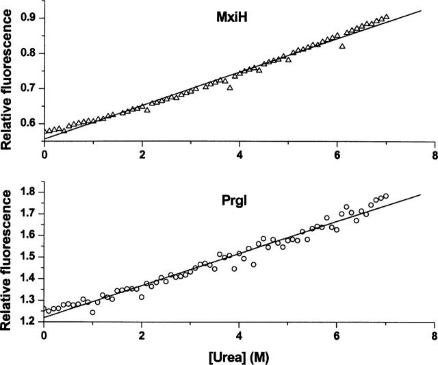
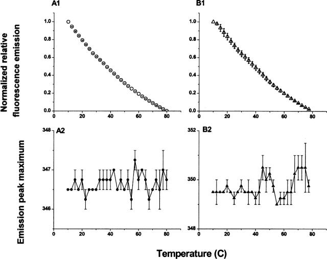
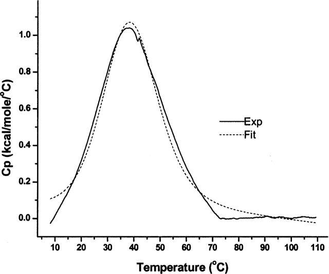
Shigella and Salmonella use similar type III secretion systems for delivering effector proteins into host cells. This secretion system consists of a base anchored in both bacterial membranes and an extracellular "needle" that forms a rod-like structure exposed on the pathogen surface. The needle is composed of multiple subunits of a single protein and makes direct contact with host cells to facilitate protein delivery. The proteins that make up the needle of Shigella and Salmonella are MxiH and PrgI, respectively. These proteins are attractive vaccine candidates because of their essential role in virulence and surface exposure. We therefore isolated, purified, and characterized the monomeric forms of MxiH and PrgI. Their far-UV circular dichroism spectra show structural similarities with hints of subtle differences in their secondary structure. Both proteins are highly helical and thermally unstable, with PrgI having a midpoint of thermal unfolding (Tm) near 37 degrees C and MxiH having a value near 42 degrees C. The two proteins also have comparable intrinsic stabilities as measured by chemically induced (urea) unfolding. MxiH, however, with a free energy of unfolding (DeltaG degrees 0,un) of 1.6 kcal/mol, is slightly more stable than PrgI (1.2 kcal/mol). The relatively low m-values obtained for the urea-induced unfolding of the proteins suggest that they undergo only a small change in solvent-accessible surface area. This argues that when MxiH and PrgI are incorporated into the needle complex, they obtain a more stable structural state through the introduction of protein-protein interactions.


Figures

Figure 1.
Figure 1.
MxiH and PrgI primary sequence alignment. Below the sequences, : designates identity and . indicates similarity.
Figure 2.
Figure 2.
Far-UV CD spectra for MxiH and PrgI at 10°C at a protein concentration of 50 μM using a 0.01-cmpath-length cell. The circles show the spectrum for MxiH, and the triangles show the spectrum of PrgI. The data shown are an average of greater than or equal to two trials.
Figure 3.
Figure 3.
Thermal unfolding of MxiH (A) and PrgI (B) as monitored by CD. The solid black traces represent unfolding, and the dashed lines, the unfolding of proteins previously heated to >90°C and then cooled.
Figure 4.
Figure 4.
MxiH thermal unfolding as monitored by UV absorbance spectroscopy. Data are derived from second derivatives of the UV spectra as a function of temperature. Peaks 1, 2, and 3 represent contributions from Phe; peaks 3, 4, and 5 from Tyr; and peaks 5 and 6 from Trp. (Peaks 3 and 5 are combination peaks.) The line in the data for peaks 4 and 5 is the fit of a two-state unfolding model to the data.
Figure 5.
Figure 5.
PrgI thermal unfolding as monitored by UV absorbance. Data are derived from second derivatives of the UV spectra at various temperatures as described in the legend to Figure 4.
Figure 6.
Figure 6.
Urea-induced unfolding of MxiH and PrgI as monitored by CD at 222 nm. Panels A1 and A2 show MxiH, and panels B1 and B2 show PrgI. The upper panels (A1,B1) show data acquired at 10°C, and the lower panels (A2,B2) show data acquired at 25°C.
Figure 7.
Figure 7.
Typical normalized fluorescence emission spectra of MxiH and PrgI (50 μM) in PBS at pH 7.0. Samples were excited at 295 nm and the emission monitored from 302 nm to 450 nm. PrgI is shown with the solid and MxiH is shown with a dashed line.
Figure 8.
Figure 8.
Urea-induced unfolding of MxiH and PrgI at 10°C as monitored by Trp fluorescence with excitation at 295 nm. The proteins were examined at 50 μM. Panels A and B represent MxiH and PrgI with slopes 0.05 and 0.07 M−1, respectively.
Figure 9.
Figure 9.
Thermal unfolding curves of MxiH and PrgI as monitored by Trp fluorescence. Panels A1 and A2 represent PrgI, while panels B1 and B2 represent MxiH. The upper panels (A1,B1) show the intensity of the fluorescence emission at 340 nm as a function of temperature. The lower panels (A2,B2) show the wavelength of the emission maximum as function of temperature.
Figure 10.
Figure 10.
PrgI DSC endotherm with buffer subtraction and baseline correction at a protein concentration of 1.82 mg/mL. A two-state unfolding model was used to fit the data and the values of Tm=38.1°C ± 0.3 and ΔH=27.9 kcal/mol ± 0.1 were found. The solid line is from a representative experiment with the dashed line showing a best fit analysis.

Similar articles

Cited by

References

    1. Allaoui, A., Sansonetti, P.J., and Parsot, C. 1992. MxiJ, a lipoprotein involved in secretion of Shigella Ipa invasions, is homologous to YscJ, a secretion factor of the Yersinia Yop proteins. J. Bacteriol. 174: 7661–7669. - PMC - PubMed
    1. Blocker, A., Jouihri, N., Larquet, E., Gounon, P., Ebel, F., Parsot, C., Sansonetti, P., and Allaoui, A. 2001. Structure and composition of the Shigella flexneri “needle complex,” a part of its type III secretion. Mol. Microbiol. 39: 652–663. - PubMed
    1. Blocker, A., Komoriya, K., and Aizawa, S. 2003. Type III secretion systems and bacterial flagella: Insights into their function from structural similarities. Proc. Natl. Acad. Sci. 100: 3027–3030. - PMC - PubMed
    1. Cordes, F.S., Komoriya, K., Larquet, E., Yang, S., Egelman, E.H., Blocker, A., and Lea, S.M. 2003. Helical structure of the needle of the type III secretion system of Shigella flexneri. J. Biol. Chem. 278: 17103–17107. - PubMed
    1. Cordes, F.S., Daniell, S., Kenjale, R., Saurya, S., Picking, W.L., Picking, W.D., Booy, F., Lea, S.M., and Blocker, A. 2005. Helical packing of needles from functionally altered Shigella type III secretions systems. J. Mol. Biol. 354: 206–211. - PubMed

Publication types

LinkOut - more resources