. 2006 Mar;15(3):647-54.
doi: 10.1110/ps.052012306.

The early history of the ubiquitin field

Affiliations

The early history of the ubiquitin field

Alexander Varshavsky. Protein Sci. 2006 Mar.

Abstract

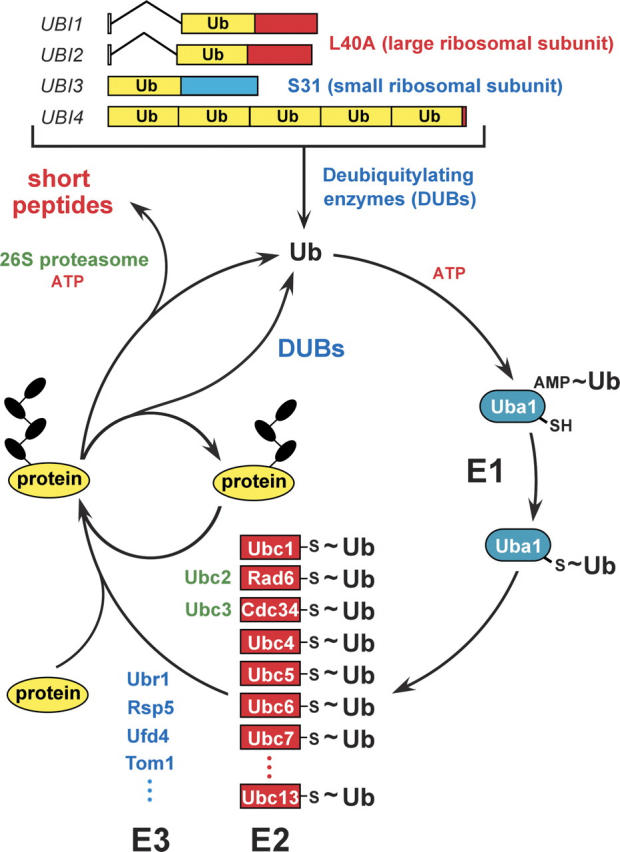
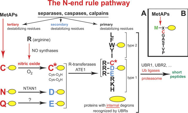
This is a personal account of the early history of ubiquitin research, by one of its protagonists. The field of ubiquitin and regulated protein degradation was created in the 1980s, largely through the complementary discoveries by the laboratory of A. Hershko (Technion, Haifa, Israel) and by my laboratory, then at MIT (Cambridge, MA). I describe the elegant insights by Hershko and his colleagues that yielded the initial understanding of ubiquitin conjugation and ubiquitin-mediated proteolysis in cell extracts, including the identification of E1, E2, and E3 enzymes. These advances were followed by a set of interconnected discoveries in my laboratory that revealed the biology of the ubiquitin system, i.e., its necessity for the protein degradation in vivo, its specific physiological functions (in the cell cycle, DNA repair, protein synthesis, transcriptional regulation, and stress responses), the source of its selectivity (specific degradation signals in short-lived proteins), and its key mechanistic attributes, such as the polyubiquitin chain and the subunit selectivity of protein degradation. The above biological (function-based) insights produced the main discovery of the physiological regulation by intracellular protein degradation. These advances caused the enormous expansion of the ubiquitin field in the 1990s. Together with the initial discovery of ubiquitin-mediated proteolysis by Hershko and coworkers, our biological discoveries in the 1980s led to a radically changed understanding of the logic of intracellular circuits, as it became clear that the control through regulated protein degradation rivals, and often surpasses in significance, the classical regulation through transcription and translation.


Figures

Figure 1.
Figure 1.
The ubiquitin system of the yeast S. cerevisiae (Varshavsky 1997, 2005). The fundamental design of this system is conserved among eukaryotes. The yeast ubiquitin genes UBI1UBI4, two of which contain introns, encode fusions of ubiquitin either to itself or to one of two ribosomal proteins. These fusions are cleaved by deubiquitylating enzymes (DUBs), yielding mature ubiquitin. Thioester bonds between ubiquitin and the active-site Cys residues of ubiquitin-specific enzymes are denoted by the “~” sign. The conjugation of ubiquitin to other proteins involves a preliminary ATP-dependent step, in which the last (Gly-76) residue of ubiquitin is joined, via a thioester bond, to a Cys residue of the ubiquitin-activating (E1) enzyme, encoded by UBA1. The activated ubiquitin is transferred to a Cys residue in one of several ubiquitin-conjugating (E2) enzymes, encoded by the UBC-family genes, and from there to a Lys residue of an ultimate acceptor protein. This last step, and the formation of a substrate-linked polyubiquitin chain (black ovals) require participation of another component, called E3, whose mechanistic functions include the recognition of a substrate’s degradation signal (degron). The names of some of the currently known yeast E3s are indicated as well. The term “ubiquitin ligase” denotes either an E2–E3 holoenzyme or its E3 component. A targeted, ubiquitylated protein is processively degraded to short peptides by the ATP-dependent 26S proteasome. (For reviews, see Varshavsky 1997; Hershko et al. 2000; Pickart 2004.)
Figure 2.
Figure 2.
(A) The N-end rule pathway in mammals (Kwon et al. 2002, Hu et al. 2005). This proteolytic pathway was the first specific pathway of the ubiquitin system to be discovered, initially in yeast (Bachmair et al. 1986; Varshavsky 1996). It is present in all eukaryotes examined, from fungi to animals and plants. Although prokaryotes lack ubiquitin conjugation and ubiquitin itself, they, too, contain the N-end rule pathway, a ubiquitin-independent version of it (Tobias et al. 1991; Shrader et al. 1993). Studies of this pathway, its mechanisms and functions, have become a major focus of my laboratory. N-terminal residues are indicated by single-letter abbreviations for amino acids. The ovals denote the rest of a protein substrate. MetAPs, methionine aminopeptidases. The “cysteine” (Cys) sector, in the upper left corner, describes the recent discovery of a nitric oxide (NO)-mediated oxidation of N-terminal Cys, with subsequent arginylation of oxidized Cys by the ATE1-encoded isoforms of Arg-tRNA-protein transferase (R-transferase) (Hu et al. 2005). This advance identified the N-end rule pathway as a new kind of NO sensor. C* denotes oxidized Cys, either Cys-sulfinic acid (CysO2[H]) or Cys-sulfonic acid (CysO3[H]). Type 1 and type 2 primary destabilizing N-terminal residues are recognized by multiple E3 ubiquitin ligases of the N-end rule pathway, including UBR1 and UBR2. Through their other substrate-binding sites, these E3 enzymes also recognize internal (non-N-terminal) degrons in other substrates of the N-end rule pathway, denoted by a larger oval. (B) MetAPs remove Met from the N terminus of a polypeptide if the residue at position 2 belongs to the set of residues shown.

Similar articles

Cited by

References

    1. Bachmair, A. and Varshavsky, A. 1989. The degradation signal in a short-lived protein. Cell 56: 1019–1032. - PubMed
    1. Bachmair, A., Finley, D., and Varshavsky, A. 1986. In vivo half-life of a protein is a function of its amino-terminal residue. Science 234: 179–186. - PubMed
    1. Bartel, B., Wünning, I., and Varshavsky, A. 1990. The recognition component of the N-end rule pathway. EMBO J. 9: 3179–3189. - PMC - PubMed
    1. Baumeister, W., Walz, J., Zühl, F., and Seemüller, E. 1998. The proteasome: Paradigm of a self-compartmentalizing protease. Cell 92: 367–380. - PubMed
    1. Chau, V., Tobias, J.W., Bachmair, A., Marriott, D., Ecker, D.J., Gonda, D.K., and Varshavsky, A. 1989. A multiubiquitin chain is confined to specific lysine in a targeted short-lived protein. Science 243: 1576–1583. - PubMed

Publication types

Personal name as subject

LinkOut - more resources