

Frontal horn cysts in normal neonates
- PMID: 16503391
- PMCID: PMC7125929
- DOI: 10.1016/j.braindev.2006.01.002
Frontal horn cysts in normal neonates
Abstract
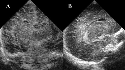
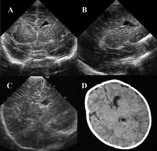
Frontal horn cysts (FHCs) are elliptical, smooth, thin-walled cysts adjacent to the tip of the anterior horns of the lateral ventricles. Among 3,545 terms or near term healthy babies who underwent cranial ultrasound examination in our hospital over a 2-year 5-month period, 18 were found to have FHCs (17 typical and one atypical; seven bilateral and 11 unilateral, of which seven were on the left and four on the right). The female to male ratio was 2:1. The incidence of FHCs in normal term babies was thus 0.5%. Six children had resolution of the cyst within 1 month, and 6 more had resolution on repeat scan from 2 to 11 months of age. Four children did not have subsequent ultrasonography to document resolution, but they had normal growth and development. Two were lost to follow up. The infant with an atypical FHC had an enlarged left frontal horn cyst with a midline shift on follow up, but he had normal development. Our study suggests that FHC may be a normal physiologic variant or a benign pathologic condition that can be expected to resolve spontaneously within a few months. It is reasonable to follow typical FHC by cranial ultrasound examinations at 1 or 2 and 6 months of age. In the case of an atypical cyst, more frequent follow up and further image studies like CT or MRI are necessary.
Figures
References
-
- Sudakoff G.S., Mitchell D.G., Stanley C., Graziani L.J. Frontal periventricular cysts on the first day of life: a one-year clinical follow-up and its significance. J Ultrasound Med. 1991;10(1):25–30. - PubMed
-
- Thun-Hohenstein L., Forster I., Kunzle C., Martin E., Boltshauser E. Transient bifrontal solitary periventricular cysts in term neonates. Neuroradiology. 1994;36(3):241–244. - PubMed
-
- Zorzi C., Angonese I. Subependymal pseudocysts in the neonate. Eur J Pediatr. 1989;148(5):462–464. - PubMed
Publication types
MeSH terms
LinkOut - more resources
Full Text Sources
Medical
