. 2006 Feb 20:5:8.
doi: 10.1186/1476-4598-5-8.

Histone deacetylase inhibitor, trichostatin A induces ubiquitin-dependent cyclin D1 degradation in MCF-7 breast cancer cells

Affiliations

Histone deacetylase inhibitor, trichostatin A induces ubiquitin-dependent cyclin D1 degradation in MCF-7 breast cancer cells

John P Alao et al. Mol Cancer. .

Abstract

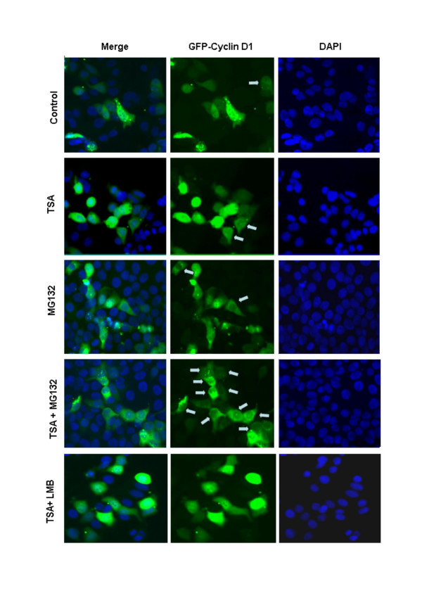
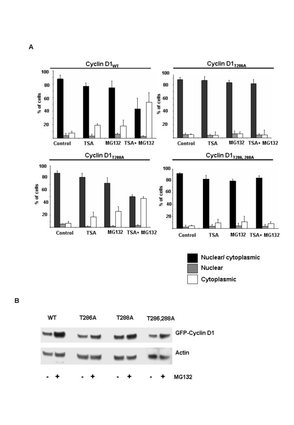
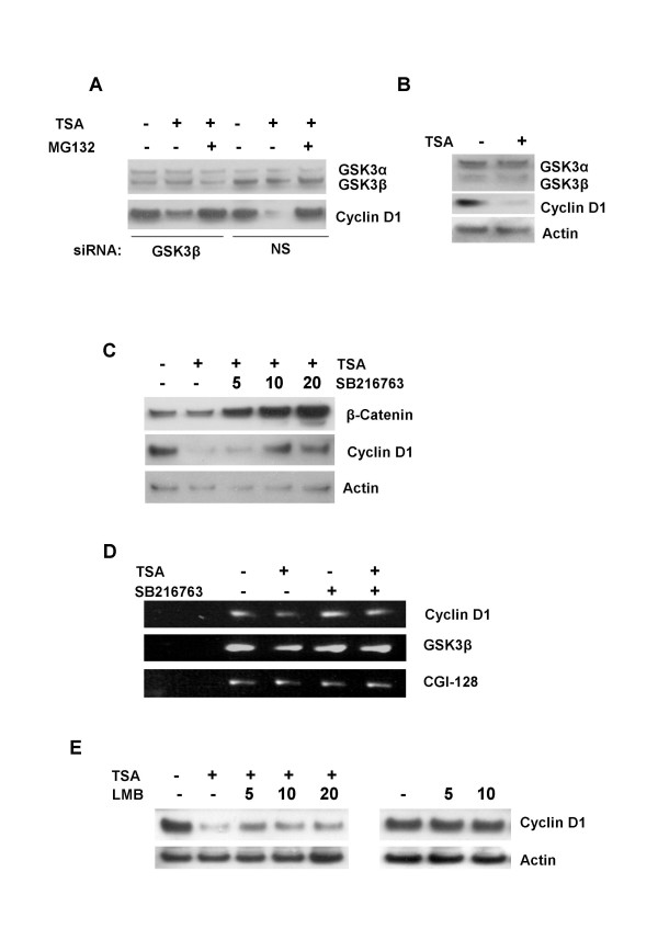
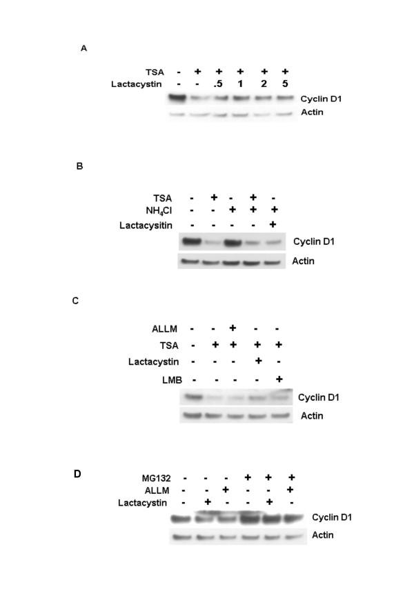
Background: Cyclin D1 is an important regulator of G1-S phase cell cycle transition and has been shown to be important for breast cancer development. GSK3beta phosphorylates cyclin D1 on Thr-286, resulting in enhanced ubiquitylation, nuclear export and degradation of the cyclin in the cytoplasm. Recent findings suggest that the development of small-molecule cyclin D1 ablative agents is of clinical relevance. We have previously shown that the histone deacetylase inhibitor trichostatin A (TSA) induces the rapid ubiquitin-dependent degradation of cyclin D1 in MCF-7 breast cancer cells prior to repression of cyclin D1 gene (CCND1) transcription. TSA treatment also resulted in accumulation of polyubiquitylated GFP-cyclin D1 species and reduced levels of the recombinant protein within the nucleus.

Results: Here we provide further evidence for TSA-induced ubiquitin-dependent degradation of cyclin D1 and demonstrate that GSK3beta-mediated nuclear export facilitates this activity. Our observations suggest that TSA treatment results in enhanced cyclin D1 degradation via the GSK3beta/CRM1-dependent nuclear export/26S proteasomal degradation pathway in MCF-7 cells.

Conclusion: We have demonstrated that rapid TSA-induced cyclin D1 degradation in MCF-7 cells requires GSK3beta-mediated Thr-286 phosphorylation and the ubiquitin-dependent 26S proteasome pathway. Drug induced cyclin D1 repression contributes to the inhibition of breast cancer cell proliferation and can sensitize cells to CDK and Akt inhibitors. In addition, anti-cyclin D1 therapy may be highly specific for treating human breast cancer. The development of potent and effective cyclin D1 ablative agents is therefore of clinical relevance. Our findings suggest that HDAC inhibitors may have therapeutic potential as small-molecule cyclin D1 ablative agents.


Figures

Figure 1
Figure 1
Effect of TSA on GFP-Cyclin D1 localization in MCF-7 cells. Cells were transfected with expression vectors encoding wild type GFP-Cyclin D1 (GFP-Cyclin D1WT). 24 h after transfection, cells were treated with TSA (1 μM) alone or in the presence of MG132 (50 μM) or leptomycin B (LMB) (10 ng /ml) for 6 h. Cells were fixed in ice cold methanol for 5 min, counterstained with DAPI and examined by fluorescence microscopy. Cells with predominantly cytoplasmic GFP-cyclin D1 are indicated by grey arrows.
Figure 2
Figure 2
Thr286 phosphorylation mediates the effect of TSA on GFP-cyclin D1 cytoplasmic accumulation A, Thr-286 but not Thr-288 is required for TSA induced cyclin D1 nuclear exclusion. MCF-7 cells were transfected with vectors encoding GFP-Cyclin D1WT, GFP-Cyclin D1T286A, GFP-Cyclin D1T288A or GFP-Cyclin D1T286,-288A for 24 h and treated as for Figure 1. GFP-Cyclin D1 localization was scored by quantitative fluorescence microscopy counting >200 cells in three separate experiments. B, Effect of MG132 on wild type and mutant GFP-Cyclin D1 protein levels. Cells were transfected as in A and then treated with MG132 (50 μM) for 6 h. Cell lysates were separated by 4–20 % SDS-PAGE and immunoblot analysis was done using antibodies against GFP and actin.
Figure 3
Figure 3
Effect of GSK3β and CRM1 inhibition on TSA induced cyclin D1 degradation. A, MCF-7 cells were transfected with or without GSK3β specific siRNA pools. 72 h after transfection, cells were treated with TSA (1 μM) alone or in the presence of MG132 (50 μM) for 6 h. Cell lysates were separated by 4–20 % SDS-PAGE and immunoblot analysis was done using antibodies against GSK3 α/β, cyclin D1 and actin. B, Effect of TSA on GSK3β levels and activity. MCF-7 cells were cultured in the absence or presence of TSA for 6 h. Immunoblot analysis was done with antibodies against GSK3, cyclin D1 and actin. C, partial inhibition of TSA induced cyclin D1 degradation by the GSK3 inhibitor SB216763. Cells were cultured for 24 h with the indicated concentrations (μM) of SB216763 and then treated for 6 h with TSA (1 μM). Immunoblot analysis was done with antibodies against β-Catenin, cyclin D1 and actin. D, Effect of SB216763 on GSK3β mRNA expression. MCF-7 cells were cultured for 24 h in the absence or presence of SB216763 (10 μM) and treated with TSA (1 μM) 12 h. Semi-quantitative RT-PCR analysis was done with primers for GSK3β, cyclin D1 and CGI-128 as a loading control. E, effect of the CRM1-dependent nuclear export inhibitor Leptomycin B (LMB) on TSA induced cyclin D1 degradation. MCF-7 cells were pretreated for 1 h with the indicated concentrations (ng/mL) of LMB and then treated with TSA (1 μM) for 6 h. Alternatively MCF-7 cells were cultured in the presence of the indicated concentrations (ng/mL) of LMB for 6 h. Immunoblot analysis was done with antibodies against cyclin D1 and actin.
Figure 4
Figure 4
Effect of lactacystin on TSA induced cyclin D1 degradation. A, MCF-7 cells were pretreated with lactacystin (10 μM) for 1 h and then treated with TSA (1 μM) for 6 h. Cell lysates were separated by 4–20 % SDS-PAGE and immunoblot analysis was done using antibodies against cyclin D1 and actin. B, effect of ammonium chloride and lactacystin on TSA induced cyclin D1 degradation. MCF-7 cells were pretreated with the indicated concentrations of ammonium chloride (10 mM) and/or lactacystin (10 μM) for 1 h and then treated with TSA (1 μM) for 6 h. Immunoblot analysis was done with antibodies against cyclin D1 and actin. C, Effects of ALLM, lactacystin and LMB on TSA induced cyclin D1 degradation. MCF-7 cells were pretreated with ALLM (100 μM), lactacystin (10 μM) or LMB (10 ng/mL) for 1 h and then treated with TSA (1 μM) for 6 h. Immunoblot analysis was done with antibodies against cyclin D1 and actin. D, Lactacystin and ALLM induced cyclin D1 loss is inhibited by MG132. MCF-7 cells were treated with lactacystin (10 μM) or ALLM (100 μM) with or without MG132 (50 μM) for 6 h. Immunoblot analysis was done with antibodies against cyclin D1 and actin.

Similar articles

Cited by

References

    1. Sherr CJ, Roberts JM. CDK inhibitors: positive and negative regulators of G1-phase progression. Genes Dev. 1999;13:1501–1512. - PubMed
    1. Sherr CJ, Roberts JM. CDK inhibitors: positive and negative regulators of G1-phase progression. Genes Dev. 1999;13:1501–1512. - PubMed
    1. Baldin V, Lukas J, Marcote MJ, Pagano M, Draetta G. Cyclin D1 is a nuclear protein required for cell cycle progression in G1. Genes Dev. 1993;7:812–821. - PubMed
    1. Guo Y, Yang K, Harwalkar J, Nye JM, Mason DR, Garrett MD, Hitomi M, Stacey DW. Phosphorylation of cyclin D1 at Thr 286 during S phase leads to its proteasomal degradation and allows efficient DNA synthesis. Oncogene. 2005;24:2599–2612. doi: 10.1038/sj.onc.1208326. - DOI - PubMed
    1. Diehl JA, Cheng M, Roussel MF, Sherr CJ. Glycogen synthase kinase-3beta regulates cyclin D1 proteolysis and subcellular localization. Genes Dev. 1998;12:3499–3511. - PMC - PubMed

Publication types

MeSH terms

Substances