. 2006 Feb 21:6:2.
doi: 10.1186/1472-6793-6-2.

Proinflammatory cytokines tumor necrosis factor-alpha and interferon-gamma modulate epithelial barrier function in Madin-Darby canine kidney cells through mitogen activated protein kinase signaling

Affiliations

Proinflammatory cytokines tumor necrosis factor-alpha and interferon-gamma modulate epithelial barrier function in Madin-Darby canine kidney cells through mitogen activated protein kinase signaling

David M Patrick et al. BMC Physiol. .

Abstract

Background: The tight junction is a dynamic structure that is regulated by a number of cellular signaling processes. Occludin, claudin-1, claudin-2 and claudin-3 are integral membrane proteins found in the tight junction of MDCK cells. These proteins are restricted to this region of the membrane by a complex array of intracellular proteins which are tethered to the cytoskeleton. Alteration of these tight junction protein complexes during pathological events leads to impaired epithelial barrier function that perturbs water and electrolyte homeostasis. We examined MDCK cell barrier function in response to challenge by the proinflammatory cytokines tumor necrosis factor-alpha (TNFalpha) and interferon-gamma (IFNgamma).

Results: Exposure of MDCK cells to TNFalpha/IFNgamma resulted in a marked sustained elevation of transepithelial electrical resistance (TER) as well as elevated paracellular permeability. We demonstrate that the combination of TNFalpha/IFNgamma at doses used in this study do not significantly induce MDCK cell apoptosis. We observed significant alterations in occludin, claudin-1 and claudin-2 protein expression, junctional localization and substantial cytoskeletal reorganization. Pharmacological inhibition of ERK1/2 and p38 signaling blocked the deleterious effects of the proinflammatory cytokines on barrier function.

Conclusion: These data strongly suggest that downstream effectors of MAP kinase signaling pathways mediate the TNFalpha/IFNgamma-induced junctional reorganization that modulates MDCK cell barrier function.


Figures

Figure 1
Figure 1
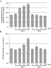
Effect of Inflammatory Cytokine Concentration on MDCK Cell Barrier Function. Lactate dehydrogenase release (Panel A) was measured in confluent MDCK cell cultures 24 hours following exposure to increasing doses of TNFα and IFNγ. Results are expressed as percent of maximal LDH release determined by incubating MDCK cells with TX-100 (2%) five minutes prior to LDH activity assay. The mean LDH is reported, error bars represent the SE, four independent experiments were assayed in duplicate. Fluorescein flux (Panel B) was measured following 24 hour treatment with increasing dose of TNFα and IFNγ. Fluorescein (50 μM) was added to the apical chamber and recovery was measured from the basal chamber after a 120 minute incubation. The mean fluorescence is reported, error bars represent the SE of four independent experiments. A one-way analysis of variance (ANOVA) was performed, multiple comparisons between control and treatments were determined with the Bonferroni post test. **Indicates statistical difference (P < 0.001) to control.
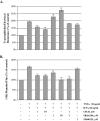
Figure 2
Figure 2
Tumor Necrosis Factor-α Elevates Transepithelial Electrical Resistance and Flux in MDCK cells. MDCK cells were treated with increasing dose of TNFα or IFNγ for 24 hours; TER and [3H]-mannitol flux were determined. Panel A reports the mean percent change in TER, TNFα produced a significant dose dependent elevation in TER whereas IFNγ had minimal effects on TER. At 24 hours post treatment [3H]-mannitol (2 μCi) was added to the apical chamber, cells were incubated at 37°C for two hours, recovery of tracer was measured in the basolateral chamber and expressed as fold change from the control group. Error bars represent the SEM, n = 3. A one-way analysis of variance (ANOVA) was performed, multiple comparisons between control and treatments were determined with the Bonferroni post test. **Indicates statistical difference (P < 0.001) to TNFα group (3 ng/ml).
Figure 3
Figure 3
Tumor Necrosis Factor-α and Interferon-γ Synergize to Elevate Flux in MDCK cells. MDCK cells were treated with increasing dose of TNFα/IFNγ for up to 72 hours; TER and [3H]-mannitol flux were determined. Panel A reports the mean TER when cells were exposed to the following conditions; media only control (formula image), TNFα/IFNγ, 3/6 ng/ml (formula image), TNFα/IFNγ, 10/20 ng/ml (formula image), and TNFα/IFNγ, 30/60 ng/ml (formula image). Panel B reports the mean [3H]-mannitol flux following 72 hour incubation with the indicated treatments. Flux is presented as the percent of apical [3H]-mannitol recovered in the basolateral chamber following 120 min. incubation. Error bars represent the SE, n = 6. A one-way analysis of variance (ANOVA) was performed, multiple comparisons between control and treatments were determined with the Bonferroni post test. **Indicates statistical difference (P < 0.001) to control.
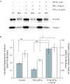
Figure 4
Figure 4
Effect of MAP Kinase Inhibition on TER and Paracellular Flux in TNFα and IFNγ-treated MDCK cells. The effect of the MAP kinase inhibitors was investigated by TER assessment and [3H]-mannitol flux determination in the presence of TNFα and IFNγ. MDCK cells were placed into one of eight treatment groups for 24 hours; control, TNFα/IFNγ alone (30/60 ng/ml) or proinflammatory cytokine with U0126 (1 and 10 μM), SB202190 (1 and 10 μM), combined U0126 and SB202190 (1 μM each) or SB600125 (1 μM). TER was assessed using the EVOM system (Panel A) then flux was determined following incubation at 37°C for two hours with [3H]-mannitol in the apical chamber (Panel B). Recovery of tracer was measured in the basolateral chamber and expressed as fold change from the control group. Exposure to TNFα/IFNγ produces a significant two-fold elevation in paracellular flux; MAP kinase inhibitors protect barrier function to varying degrees. Error bars represent the mean ± SE of four independent experiments. ANOVA was performed, multiple comparisons between all treatments were determined with the Tukey-HSD post test. **Indicates statistically difference (P < 0.01) to the TNFα/IFNγ group.
Figure 5
Figure 5
Effect of Proinflammatory Cytokine Concentration on MDCK Cell Tight Junction Proteins. Representative immunoblots of occludin, claudin-1, claudin-2 and claudin-3 total protein from confluent MDCK cell cultures twenty-four hours following exposure to increasing doses of TNFα and IFNγ (Panel A). Immunoblots were subjected to densitometric analysis; results are reported as percent of control to examine the effect of proinflammatory cytokines on tight junction protein expression (Panel B). Dashed horizontal lines represent control level of expression (100%), error bars represent the mean ± SE of eight independent experiments. ANOVA was performed, multiple comparisons between control and treatments were determined with the Bonferroni post test. **Indicates statistically difference (P < 0.01) to control.
Figure 6
Figure 6
ERK1/2 Inhibition Reverses Cytokine-induced Alterations in Tight Junction Protein Distribution. Representative immunoblots of occludin and claudin-1 using Triton X-100 soluble and insoluble (SDS) fractions from confluent MDCK cell cultures treated for 24 hours in the indicated conditions (Panel A). The effect of the MEK inhibitor (U0126, 1 μM) was added fifteen minute prior to addition of proinflammatory cytokines. Densitometic analyses were performed, occludin (filled bars) and claudin-1 (open bars), ratio of SDS to TX-100 intensity was reported. Error bars represent the mean ± SE of four independent experiments. ANOVA was performed, multiple comparisons between all treatments were determined with the Tukey-HSD post test. **Indicates statistically difference (P < 0.05) to the control group, ***indicates a significant difference to the TNFα/IFNγ group (P < 0.05).
Figure 7
Figure 7
Aberrant Localization of Tight Junction Proteins and Cytoskeletal Reorganization induced by Proinflammatory Cytokines in MDCK Cells. Immunofluorescence microscopy was used to capture digital images of MDCK cells grown to confluency on glass coverslips. Images A, D, G, J and M show control occludin, claudin-1, claudin-2, claudin-3 and actin staining respectively. Images B, E, H, K and N demonstrate occludin, claudin-1, claudin-2, claudin-3 and actin staining following twenty-four treatment with TNFα/IFNγ (10 and 20 ng/ml). Cells were treated TNFα/IFNγ for a 24 hour interval in the presence the ERK1/2 inhibitor U0126, 1 μM, representative images C, F, I, L and O were stained for occludin, claudin-1, claudin-2, claudin-3 and actin respectively. All images were captured on a Nikon 2000E microscope using a 60X oil-immersion lens, the calibration bar represents 25 μm.

Similar articles

Cited by

References

    1. Anderson JM, Van Itallie CM. Tight junctions and the molecular basis for regulation of paracellular permeability. Am J Physiol. 1995;269:G467–75. - PubMed
    1. Furuse M, Hirase T, Itoh M, Nagafuchi A, Yonemura S, Tsukita S. Occludin: a novel integral membrane protein localizing at tight junctions. J Cell Biol. 1993;123:1777–1788. doi: 10.1083/jcb.123.6.1777. - DOI - PMC - PubMed
    1. Martin-Padura I, Lostaglio S, Schneemann M, Williams L, Romano M, Fruscella P, Panzeri C, Stoppacciaro A, Ruco L, Villa A, Simmons D, Dejana E. Junctional adhesion molecule, a novel member of the immunoglobulin superfamily that distributes at intercellular junctions and modulates monocyte transmigration. J Cell Biol. 1998;142:117–127. doi: 10.1083/jcb.142.1.117. - DOI - PMC - PubMed
    1. Furuse M, Sasaki H, Fujimoto K, Tsukita S. A single gene product, claudin-1 or -2, reconstitutes tight junction strands and recruits occludin in fibroblasts. J Cell Biol. 1998;143:391–401. doi: 10.1083/jcb.143.2.391. - DOI - PMC - PubMed
    1. Mitic LL, Van Itallie CM, Anderson JM. Molecular physiology and pathophysiology of tight junctions I. Tight junction structure and function: lessons from mutant animals and proteins. Am J Physiol Gastrointest Liver Physiol. 2000;279:G250–4. - PubMed

Publication types

MeSH terms

LinkOut - more resources