

Dexamethasone protection from TNF-alpha-induced cell death in MCF-7 cells requires NF-kappaB and is independent from AKT
- PMID: 16504042
- PMCID: PMC1395311
- DOI: 10.1186/1471-2121-7-9
Dexamethasone protection from TNF-alpha-induced cell death in MCF-7 cells requires NF-kappaB and is independent from AKT
Abstract
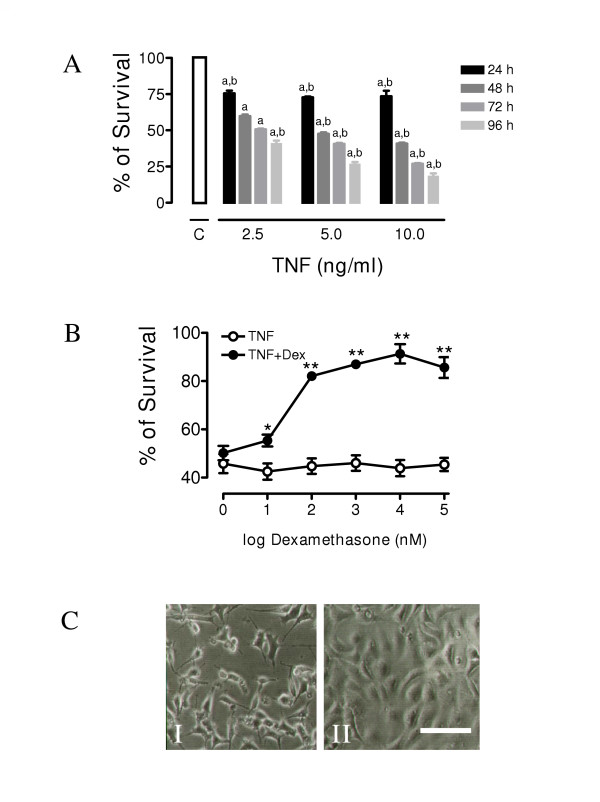
Background: The biochemical bases for hormone dependence in breast cancer have been recognized as an important element in tumor resistance, proliferation and metastasis. On this respect, dexamethasone (Dex) dependent protection against TNF-alpha-mediated cell death in the MCF-7 cell line has been demonstrated to be a useful model for the study of this type of cancer. Recently, cytoplasmic signaling induced by steroid receptors has been described, such as the activation of the PI3K/Akt and NF-kappaB pathways. We evaluated their possible participation in the Dex-dependent protection against TNF-alpha-mediated cell death.
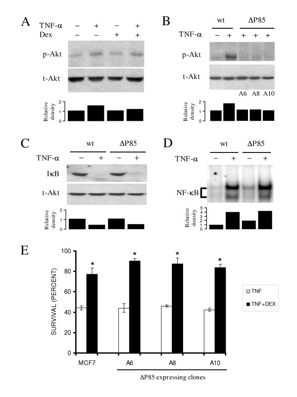
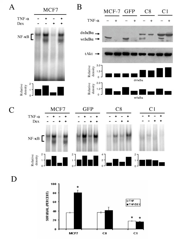
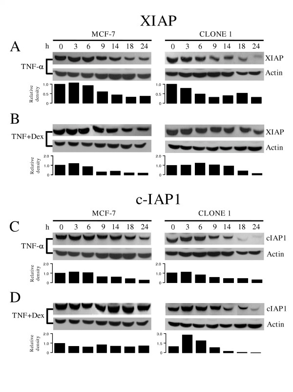
Results: Cellular cultures of the MCF-7 cell line were exposed to either, TNF-alpha or TNF-alpha and Dex, and cell viability was evaluated. Next, negative dominants of PI3K and IkappaB-alpha, designed to block the PI3K/Akt and NF-kappaB pathways, respectively, were transfected and selection and evaluation of several clones overexpressing the mutants were examined. Also, correlation with inhibitor of apoptosis proteins (IAPs) expression was examined. Independent inhibition of these two pathways allowed us to test their participation in Dex-dependent protection against TNF-alpha-cytotoxicity in MCF-7 cells. Expression of the PI3K dominant negative mutant did not alter the protection conferred by Dex against TNF-alpha mediated cell death. Contrariwise, clones expressing the IkappaB-alpha dominant negative mutant lost the Dex-conferred protection against TNF-alpha. In these clones degradation of c-IAP was accelerated, while that of XIAP was remained unaffected.
Conclusion: NF-kappaB, but not PI3K/Akt activation, is required for the Dex protective effect against TNF-alpha-mediated cell death, and correlates with lack of degradation of the anti-apoptotic protein c-IAP1.
Figures
Similar articles
-
NF-kappaB activation but not PI3K/Akt is required for dexamethasone dependent protection against TNF-alpha cytotoxicity in L929 cells.FEBS Lett. 2005 Jul 18;579(18):3947-52. doi: 10.1016/j.febslet.2005.05.081. FEBS Lett. 2005. PMID: 16000198
-
Dexamethasone suppresses interleukin-1beta-induced human beta-defensin 2 mRNA expression: involvement of p38 MAPK, JNK, MKP-1, and NF-kappaB transcriptional factor in A549 cells.FEMS Immunol Med Microbiol. 2007 Oct;51(1):171-84. doi: 10.1111/j.1574-695X.2007.00293.x. Epub 2007 Jul 23. FEMS Immunol Med Microbiol. 2007. PMID: 17645739
-
MAP kinase-dependent, NF-kappaB-independent regulation of inhibitor of apoptosis protein genes by TNF-alpha.J Cell Physiol. 2007 Mar;210(3):703-10. doi: 10.1002/jcp.20881. J Cell Physiol. 2007. PMID: 17133355
-
[IAPs: a central element in the NF-κB activating signaling pathway].Med Sci (Paris). 2012 Jan;28(1):69-75. doi: 10.1051/medsci/2012281019. Epub 2012 Jan 27. Med Sci (Paris). 2012. PMID: 22289833 Review. French.
-
(Un)expected roles of c-IAPs in apoptotic and NFkappaB signaling pathways.Cell Cycle. 2008 Jun 1;7(11):1511-21. doi: 10.4161/cc.7.11.5959. Epub 2008 Mar 16. Cell Cycle. 2008. PMID: 18469528 Review.
Cited by
-
Genomic and non-genomic effects of glucocorticoids: implications for breast cancer.Int J Clin Exp Pathol. 2015 Jan 1;8(1):1-10. eCollection 2015. Int J Clin Exp Pathol. 2015. PMID: 25755688 Free PMC article. Review.
-
IL-1β Promotes a New Function of DNase I as a Transcription Factor for the Fas Receptor Gene.Front Cell Dev Biol. 2018 Feb 6;6:7. doi: 10.3389/fcell.2018.00007. eCollection 2018. Front Cell Dev Biol. 2018. PMID: 29468159 Free PMC article.
-
The glucocorticoid receptor signalling in breast cancer.J Cell Mol Med. 2008 Jan-Feb;12(1):145-63. doi: 10.1111/j.1582-4934.2007.00177.x. Epub 2007 Dec 5. J Cell Mol Med. 2008. PMID: 18053085 Free PMC article. Review.
-
Extracellular vesicles released by ALL patients contain HNE-adducted proteins: Implications of collateral damage.Free Radic Biol Med. 2025 Feb 1;227:312-321. doi: 10.1016/j.freeradbiomed.2024.12.006. Epub 2024 Dec 4. Free Radic Biol Med. 2025. PMID: 39643137
-
A biphasic effect of TNF-α in regulation of the Keap1/Nrf2 pathway in cardiomyocytes.Redox Biol. 2016 Oct;9:77-89. doi: 10.1016/j.redox.2016.06.004. Epub 2016 Jun 27. Redox Biol. 2016. PMID: 27423013 Free PMC article.
References
-
- Muti P. The role of endogenous hormones in the etiology and prevention of breast cancer: the epidemiological evidence. Recent Results Cancer Res. 2005;166:245–256. - PubMed
MeSH terms
Substances
LinkOut - more resources
Full Text Sources
Other Literature Sources
Research Materials
Miscellaneous
