

Ventricular volume and dementia progression in the Cardiovascular Health Study
- PMID: 16504345
- PMCID: PMC2866509
- DOI: 10.1016/j.neurobiolaging.2006.01.006
Ventricular volume and dementia progression in the Cardiovascular Health Study
Abstract
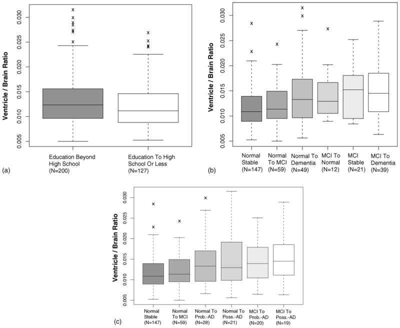
Elevated cerebral ventricular volume may be associated with dementia risk and progression. A fully-automated technique that agreed highly with radiological readings was used to estimate lateral ventricle volume on MR scans done at baseline in 1997-99 of 377 subjects in the Cardiovascular Health Study (CHS) from the Pittsburgh Center. 327 subjects were normal or diagnosed with mild cognitive impairment (MCI) at baseline and were evaluated 4 years later. Baseline ventricular volume was analyzed in multivariate models with age, gender, education level, presence and incidence of cerebral infarcts, and dementia category (normal, MCI, or dementia) at baseline and follow-up as fixed effects. Ventricular volume at baseline was significantly higher among subjects normal at baseline and demented 4 years later. Age, gender, education level, and dementia progression were significant factors affecting ventricular volume. Ventricular volume was higher in dementia compared to MCI, higher in MCI compared to controls, and higher in Possible-Alzheimer's-disease (AD) dementia compared to Probable-AD. Larger ventricles in healthy subjects may indicate susceptibility to, or progression of, dementia-related pathology.
Conflict of interest statement
Figures


References
-
- Adak S, Illouz K, Gorman W, Tandon R, Zimmerman E, Guariglia R, et al. Predicting the rate of cognitive decline in aging and early Alzheimer disease. Neurology. 2004;63(1):108–14. - PubMed
-
- Anstey K, Maller J. The role of volumetric MRI in understanding mild cognitive impairment and similar classifications. Aging Ment Health. 2003;7(4):238–50. - PubMed
-
- Breteler M, van Amerongen N, van Swieten J, Claus J, Grobbee D, van Gijn J, et al. Cognitive correlates of ventricular enlargement and cerebral white matter lesions on magnetic resonance imaging. The Rotterdam study Stroke. 1994;25(6):1109–15. - PubMed
Publication types
MeSH terms
Grants and funding
- AG15928/AG/NIA NIH HHS/United States
- AG016570/AG/NIA NIH HHS/United States
- R01 AG015928/AG/NIA NIH HHS/United States
- R21 RR019771/RR/NCRR NIH HHS/United States
- R56 AG020098/AG/NIA NIH HHS/United States
- AG05133/AG/NIA NIH HHS/United States
- U54 RR021813/RR/NCRR NIH HHS/United States
- RR021813/RR/NCRR NIH HHS/United States
- P50 CA128301/CA/NCI NIH HHS/United States
- NS07391/NS/NINDS NIH HHS/United States
- R21 EB001561/EB/NIBIB NIH HHS/United States
- T32 NS007391/NS/NINDS NIH HHS/United States
- MH01077/MH/NIMH NIH HHS/United States
- K24 MH064625/MH/NIMH NIH HHS/United States
- RR019771/RR/NCRR NIH HHS/United States
- P50 AG016570/AG/NIA NIH HHS/United States
- DA015900-01/DA/NIDA NIH HHS/United States
- P50 AG005133/AG/NIA NIH HHS/United States
- P41 RR013642/RR/NCRR NIH HHS/United States
- EB001561/EB/NIBIB NIH HHS/United States
- R01 AG020098/AG/NIA NIH HHS/United States
- R21 DA015900/DA/NIDA NIH HHS/United States
- AG20098/AG/NIA NIH HHS/United States
- MH064625/MH/NIMH NIH HHS/United States
