

Osteopontin expression and serum levels in metastatic uveal melanoma: a pilot study
- PMID: 16505010
- PMCID: PMC1414783
- DOI: 10.1167/iovs.05-0422
Osteopontin expression and serum levels in metastatic uveal melanoma: a pilot study
Abstract
Purpose: This was a pilot study conducted to examine the expression of osteopontin in uveal melanoma and to determine whether serum osteopontin can be used in detecting metastatic uveal melanoma.
Methods: Osteopontin mRNA was measured in three uveal melanoma cell lines of various invasive potential by real-time PCR. Tissue sections of primary and metastatic uveal melanomas were stained for osteopontin. Serum osteopontin levels were measured by ELISA assays in 15 patients with metastatic uveal melanoma and in 37 patients who were disease-free for at least 10 years after treatment of the primary tumor. Paired serum samples drawn from eight patients before and after development of metastasis were analyzed.
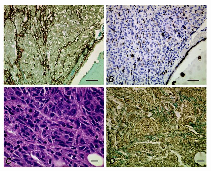
Results: By real-time PCR, highly invasive primary and metastatic uveal melanoma cells expressed 6- and 250-fold excess osteopontin mRNA, respectively, compared with poorly invasive primary uveal melanoma cells. Tissue sections of primary uveal melanomas lacking looping vasculogenic mimicry patterns either did not stain for osteopontin or exhibited weak, diffuse staining. In primary melanomas containing looping vasculogenic mimicry patterns, strong osteopontin staining was detected in the tumor periphery where patterns were located. Diffuse strong expression of osteopontin was detected in eight samples of uveal melanomas metastatic to the liver. Serum osteopontin levels were significantly higher in patients with metastatic uveal melanoma than in patients who had been disease free for at least 10 years after treatment (P = 0.0001) or in age-matched control subjects. Serum osteopontin levels were significantly higher (P = 0.008) after metastasis than before the detection of metastasis in eight patients. When a cutoff of 10 ng/mL was used, the sensitivity and specificity of serum osteopontin in detecting metastatic melanoma was 87.5%, and the area under the receiver operator characteristic curve was 96%.
Conclusions: Osteopontin is expressed diffusely in tissue sections of hepatic metastases from uveal melanoma, and increased serum osteopontin levels correlate with melanoma metastasis to the liver with high specificity and sensitivity.
Figures
References
-
- McLean IW. The biology of haematogenous metastasis in human uveal malignant melanoma. Virchows Arch A Pathol Anat. 1993;422:433–437. - PubMed
-
- Donoso LA, Shields JA, Augsburger JA, Orth DH, Johnson P. Metastatic uveal melanoma: diffuse hepatic metastasis in a patient with concurrent normal serum enzyme levels and liver scan. Arch Ophthalmol. 1985;103:758. - PubMed
-
- Eskelin S, Pyrhonen S, Summanen P, Prause JU, Kivela T. Screening for metastatic malignant melanoma of the uvea revisited. Cancer. 1999;85:1151–1159. - PubMed
-
- Kaiserman I, Amer R, Pe'er J. Liver function tests in metastatic uveal melanoma. Am J Ophthalmol. 2004;137:236–243. - PubMed
-
- Wai PY, Kuo PC. The role of Osteopontin in tumor metastasis. J Surg Res. 2004;121:228–241. - PubMed
Publication types
MeSH terms
Substances
Grants and funding
LinkOut - more resources
Full Text Sources
Other Literature Sources
Medical
Research Materials
Miscellaneous
